6COZ

Human CLC-1 chloride ion channel, C-terminal cytosolic domain


Experimental Data Snapshot

  • Method: ELECTRON MICROSCOPY
  • Resolution: 3.36 Å
  • Aggregation State: PARTICLE 
  • Reconstruction Method: SINGLE PARTICLE 

wwPDB Validation   3D Report Full Report


This is version 1.2 of the entry. See complete history


Literature

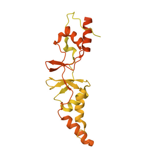
Structure of the CLC-1 chloride channel fromHomo sapiens.

Park, E.MacKinnon, R.

(2018) Elife 7

  • DOI: https://doi.org/10.7554/eLife.36629
  • Primary Citation of Related Structures:  
    6COY, 6COZ

  • PubMed Abstract: 

    CLC channels mediate passive Cl - conduction, while CLC transporters mediate active Cl - transport coupled to H + transport in the opposite direction. The distinction between CLC-0/1/2 channels and CLC transporters seems undetectable by amino acid sequence. To understand why they are different functionally we determined the structure of the human CLC-1 channel. Its 'glutamate gate' residue, known to mediate proton transfer in CLC transporters, adopts a location in the structure that appears to preclude it from its transport function. Furthermore, smaller side chains produce a wider pore near the intracellular surface, potentially reducing a kinetic barrier for Cl - conduction. When the corresponding residues are mutated in a transporter, it is converted to a channel. Finally, Cl - at key sites in the pore appear to interact with reduced affinity compared to transporters. Thus, subtle differences in glutamate gate conformation, internal pore diameter and Cl - affinity distinguish CLC channels and transporters.


  • Organizational Affiliation

    Laboratory of Molecular Neurobiology and Biophysics, Howard Hughes Medical Institute, The Rockefeller University, New York, United States.


Macromolecules
Find similar proteins by:  (by identity cutoff)  |  3D Structure
Entity ID: 1
MoleculeChains Sequence LengthOrganismDetailsImage
Chloride channel protein 1
A, B
988Homo sapiensMutation(s): 0 
Gene Names: CLCN1CLC1
Membrane Entity: Yes 
UniProt & NIH Common Fund Data Resources
Find proteins for P35523 (Homo sapiens)
Explore P35523 
Go to UniProtKB:  P35523
PHAROS:  P35523
GTEx:  ENSG00000188037 
Entity Groups  
Sequence Clusters30% Identity50% Identity70% Identity90% Identity95% Identity100% Identity
UniProt GroupP35523
Sequence Annotations
Expand
  • Reference Sequence
Experimental Data & Validation

Experimental Data

  • Method: ELECTRON MICROSCOPY
  • Resolution: 3.36 Å
  • Aggregation State: PARTICLE 
  • Reconstruction Method: SINGLE PARTICLE 
EM Software:
TaskSoftware PackageVersion
RECONSTRUCTIONRELION
MODEL REFINEMENTRosetta

Structure Validation

View Full Validation Report


Entry History & Funding Information

Deposition Data


Funding OrganizationLocationGrant Number
Howard Hughes Medical Institute (HHMI)United States--

Revision History  (Full details and data files)

  • Version 1.0: 2018-06-13
    Type: Initial release
  • Version 1.1: 2019-11-20
    Changes: Author supporting evidence
  • Version 1.2: 2024-03-13
    Changes: Data collection, Database references