6BUV

Structure of Mycobacterium tuberculosis NadD in complex with inhibitor [(1~{R},2~{R},5~{S})-5-methyl-2-propan-2-yl-cyclohexyl] 2-[3-methyl-2-(phenoxymethyl)benzimidazol-1-yl]ethanoate


Experimental Data Snapshot

  • Method: X-RAY DIFFRACTION
  • Resolution: 1.86 Å
  • R-Value Free: 0.192 
  • R-Value Work: 0.171 
  • R-Value Observed: 0.172 

wwPDB Validation   3D Report Full Report


Ligand Structure Quality Assessment 


This is version 2.0 of the entry. See complete history


Literature

Novel Antimycobacterial Compounds Suppress NAD Biogenesis by Targeting a Unique Pocket of NaMN Adenylyltransferase.

Osterman, A.L.Rodionova, I.Li, X.Sergienko, E.Ma, C.T.Catanzaro, A.Pettigrove, M.E.Reed, R.W.Gupta, R.Rohde, K.H.Korotkov, K.V.Sorci, L.

(2019) ACS Chem Biol 14: 949-958

  • DOI: https://doi.org/10.1021/acschembio.9b00124
  • Primary Citation of Related Structures:  
    6BUV

  • PubMed Abstract: 

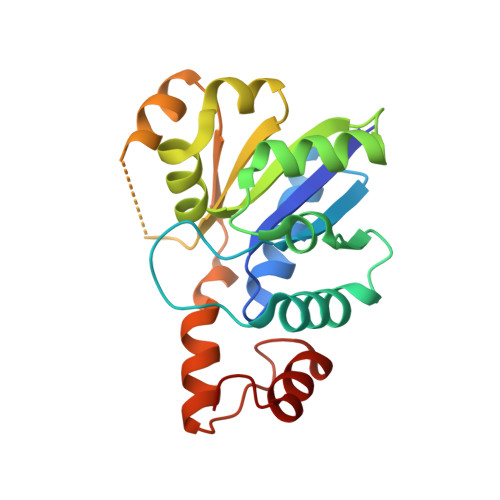
    Conventional treatments to combat the tuberculosis (TB) epidemic are falling short, thus encouraging the search for novel antitubercular drugs acting on unexplored molecular targets. Several whole-cell phenotypic screenings have delivered bioactive compounds with potent antitubercular activity. However, their cellular target and mechanism of action remain largely unknown. Further evaluation of these compounds may include their screening in search for known antitubercular drug targets hits. Here, a collection of nearly 1400 mycobactericidal compounds was screened against Mycobacterium tuberculosis NaMN adenylyltransferase ( MtNadD), a key enzyme in the biogenesis of NAD cofactor that was recently validated as a new drug target for dormant and active tuberculosis. We found three chemotypes that efficiently inhibit MtNadD in the low micromolar range in vitro. SAR and cheminformatics studies of commercially available analogues point to a series of benzimidazolium derivatives, here named N2, with bactericidal activity on different mycobacteria, including M. abscessus, multidrug-resistant M. tuberculosis, and dormant M. smegmatis. The on-target activity was supported by the increased resistance of an M. smegmatis strain overexpressing the target and by a rapid decline in NAD(H) levels. A cocrystal structure of MtNadD with N2-8 inhibitor reveals that the binding of the inhibitor induced the formation of a new quaternary structure, a dimer-of-dimers where two copies of the inhibitor occupy symmetrical positions in the dimer interface, thus paving the way for the development of a new generation of selective MtNadD bioactive inhibitors. All these results strongly suggest that pharmacological inhibition of MtNadD is an effective strategy to combat dormant and resistant Mtb strains.


  • Organizational Affiliation

    Sanford Burnham Prebys Medical Discovery Institute , La Jolla , California 92037 , United States.


Macromolecules
Find similar proteins by:  (by identity cutoff)  |  3D Structure
Entity ID: 1
MoleculeChains Sequence LengthOrganismDetailsImage
nicotinate mononucleotide adenylyltransferase NadD
A, B
201Mycobacterium tuberculosis H37RvMutation(s): 2 
Gene Names: nadDRv2421cMTCY428.26
EC: 2.7.7.18
UniProt
Find proteins for P9WJJ5 (Mycobacterium tuberculosis (strain ATCC 25618 / H37Rv))
Explore P9WJJ5 
Go to UniProtKB:  P9WJJ5
Entity Groups  
Sequence Clusters30% Identity50% Identity70% Identity90% Identity95% Identity100% Identity
UniProt GroupP9WJJ5
Sequence Annotations
Expand
  • Reference Sequence
Small Molecules
Ligands 3 Unique
IDChains Name / Formula / InChI Key2D Diagram3D Interactions
E9A (Subject of Investigation/LOI)
Query on E9A

Download Ideal Coordinates CCD File 
G [auth B]1-methyl-3-(2-{[(1R,2R,5S)-5-methyl-2-(propan-2-yl)cyclohexyl]oxy}-2-oxoethyl)-2-(phenoxymethyl)-1H-1,3-benzimidazol-3-ium
C27 H35 N2 O3
HACNGQQLOZYIEB-NIRIFSCTSA-N
CL
Query on CL

Download Ideal Coordinates CCD File 
D [auth A],
E [auth A],
F [auth A],
H [auth B],
I [auth B]
CHLORIDE ION
Cl
VEXZGXHMUGYJMC-UHFFFAOYSA-M
NA
Query on NA

Download Ideal Coordinates CCD File 
C [auth A]SODIUM ION
Na
FKNQFGJONOIPTF-UHFFFAOYSA-N
Experimental Data & Validation

Experimental Data

  • Method: X-RAY DIFFRACTION
  • Resolution: 1.86 Å
  • R-Value Free: 0.192 
  • R-Value Work: 0.171 
  • R-Value Observed: 0.172 
  • Space Group: H 3 2
Unit Cell:
Length ( Å )Angle ( ˚ )
a = 163.73α = 90
b = 163.73β = 90
c = 153.65γ = 120
Software Package:
Software NamePurpose
XSCALEdata scaling
PHASERphasing
PHENIXrefinement
PDB_EXTRACTdata extraction
XDSdata reduction

Structure Validation

View Full Validation Report


Ligand Structure Quality Assessment 


Entry History & Funding Information

Deposition Data


Funding OrganizationLocationGrant Number
National Institutes of Health/National Institute Of Allergy and Infectious Diseases (NIH/NIAID)United States1R03AI117361
National Institutes of Health/National Institute of General Medical Sciences (NIH/NIGMS)United StatesP20GM103486
National Institutes of Health/National Institute of General Medical Sciences (NIH/NIGMS)United StatesP30GM110787

Revision History  (Full details and data files)

  • Version 1.0: 2018-12-12
    Type: Initial release
  • Version 1.1: 2019-02-20
    Changes: Author supporting evidence, Data collection
  • Version 1.2: 2019-04-24
    Changes: Data collection, Database references
  • Version 1.3: 2019-05-29
    Changes: Data collection, Database references
  • Version 1.4: 2019-12-18
    Changes: Author supporting evidence
  • Version 1.5: 2023-10-04
    Changes: Data collection, Database references, Derived calculations, Refinement description
  • Version 2.0: 2024-05-01
    Changes: Derived calculations, Non-polymer description, Structure summary