6BQ8

Joint X-ray/neutron structure of PKG II CNB-B domain in complex with 8-pCPT-cGMP


Experimental Data Snapshot

  • Method: X-RAY DIFFRACTION
  • Resolution: 2.00 Å
  • R-Value Free: 0.245 
  • R-Value Work: 0.199 

  • Method: NEUTRON DIFFRACTION
  • Resolution: 2.20 Å
  • R-Value Free: 0.288 
  • R-Value Work: 0.232 

wwPDB Validation   3D Report Full Report


Ligand Structure Quality Assessment 


This is version 3.1 of the entry. See complete history


Literature

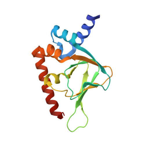
Neutron Crystallography Detects Differences in Protein Dynamics: Structure of the PKG II Cyclic Nucleotide Binding Domain in Complex with an Activator.

Gerlits, O.Campbell, J.C.Blakeley, M.P.Kim, C.Kovalevsky, A.

(2018) Biochemistry 57: 1833-1837

  • DOI: https://doi.org/10.1021/acs.biochem.8b00010
  • Primary Citation of Related Structures:  
    6BQ8

  • PubMed Abstract: 

    As one of the main receptors of a second messenger, cGMP, cGMP-dependent protein kinase (PKG) isoforms I and II regulate distinct physiological processes. The design of isoform-specific activators is thus of great biomedical importance and requires detailed structural information about PKG isoforms bound with activators, including accurate positions of hydrogen atoms and a description of the hydrogen bonding and water architecture. Here, we determined a 2.2 Å room-temperature joint X-ray/neutron (XN) structure of the human PKG II carboxyl cyclic nucleotide binding (CNB-B) domain bound with a potent PKG II activator, 8-pCPT-cGMP. The XN structure directly visualizes intermolecular interactions and reveals changes in hydrogen bonding patterns upon comparison to the X-ray structure determined at cryo-temperatures. Comparative analysis of the backbone hydrogen/deuterium exchange patterns in PKG II:8-pCPT-cGMP and previously reported PKG Iβ:cGMP XN structures suggests that the ability of these agonists to activate PKG is related to how effectively they quench dynamics of the cyclic nucleotide binding pocket and the surrounding regions.


  • Organizational Affiliation

    Bredesen Center , University of Tennessee , Knoxville , Tennessee 37996 , United States.


Macromolecules
Find similar proteins by:  (by identity cutoff)  |  3D Structure
Entity ID: 1
MoleculeChains Sequence LengthOrganismDetailsImage
cGMP-dependent protein kinase 2152Homo sapiensMutation(s): 0 
Gene Names: PRKG2PRKGR2
EC: 2.7.11.12
UniProt & NIH Common Fund Data Resources
Find proteins for Q13237 (Homo sapiens)
Explore Q13237 
Go to UniProtKB:  Q13237
PHAROS:  Q13237
GTEx:  ENSG00000138669 
Entity Groups  
Sequence Clusters30% Identity50% Identity70% Identity90% Identity95% Identity100% Identity
UniProt GroupQ13237
Sequence Annotations
Expand
  • Reference Sequence
Small Molecules
Ligands 3 Unique
IDChains Name / Formula / InChI Key2D Diagram3D Interactions
WNU
Query on WNU

Download Ideal Coordinates CCD File 
B [auth A]2-(~2~H_2_)amino-8-[(4-chlorophenyl)sulfanyl]-9-[(2S,4aR,6R,7R,7aS)-2-hydroxy-7-(~2~H)hydroxy-2-oxotetrahydro-2H,4H-2lambda~5~-furo[3,2-d][1,3,2]dioxaphosphinin-6-yl](~2~H)-1,9-dihydro-6H-purin-6-one
C16 H15 Cl N5 O7 P S
ZDJHIEHUVPCEDK-IDTAVKCVSA-N
MPD
Query on MPD

Download Ideal Coordinates CCD File 
D [auth A](4S)-2-METHYL-2,4-PENTANEDIOL
C6 H14 O2
SVTBMSDMJJWYQN-YFKPBYRVSA-N
SR
Query on SR

Download Ideal Coordinates CCD File 
C [auth A]STRONTIUM ION
Sr
PWYYWQHXAPXYMF-UHFFFAOYSA-N
Binding Affinity Annotations 
IDSourceBinding Affinity
6FW BindingDB:  6BQ8 EC50: min: 1, max: 257 (nM) from 8 assay(s)
Experimental Data & Validation

Experimental Data

  • Method: X-RAY DIFFRACTION
  • Resolution: 2.00 Å
  • R-Value Free: 0.245 
  • R-Value Work: 0.199 
  • Space Group: P 21 21 21
  • Method: NEUTRON DIFFRACTION
  • Resolution: 2.20 Å
  • R-Value Free: 0.288 
  • R-Value Work: 0.232 
  • Space Group: P 21 21 21
Unit Cell:
Length ( Å )Angle ( ˚ )
a = 43.65α = 90
b = 51.3β = 90
c = 68.2γ = 90
Software Package:
Software NamePurpose
nCNSrefinement
LAUEGENdata reduction
LSCALEdata scaling
PHASERphasing

Structure Validation

View Full Validation Report


Ligand Structure Quality Assessment 


Entry History & Funding Information

Deposition Data


Funding OrganizationLocationGrant Number
National Institutes of Health/National Institute of General Medical Sciences (NIH/NIGMS)United StatesR01 GM090161

Revision History  (Full details and data files)

  • Version 1.0: 2018-03-21
    Type: Initial release
  • Version 1.1: 2018-04-11
    Changes: Data collection, Database references
  • Version 1.2: 2019-02-20
    Changes: Author supporting evidence, Data collection
  • Version 1.3: 2020-01-01
    Changes: Author supporting evidence
  • Version 2.0: 2023-05-10
    Changes: Atomic model, Data collection, Database references, Derived calculations, Non-polymer description, Structure summary
  • Version 3.0: 2023-05-17
    Changes: Non-polymer description
  • Version 3.1: 2024-04-03
    Changes: Data collection, Refinement description