5YHL

Crystal structure of the human prostaglandin E receptor EP4 in complex with Fab and an antagonist Br-derivative


Experimental Data Snapshot

  • Method: X-RAY DIFFRACTION
  • Resolution: 4.20 Å
  • R-Value Free: 0.359 
  • R-Value Work: 0.333 
  • R-Value Observed: 0.334 

wwPDB Validation   3D Report Full Report


Ligand Structure Quality Assessment 


This is version 1.5 of the entry. See complete history


Literature

Ligand binding to human prostaglandin E receptor EP4at the lipid-bilayer interface.

Toyoda, Y.Morimoto, K.Suno, R.Horita, S.Yamashita, K.Hirata, K.Sekiguchi, Y.Yasuda, S.Shiroishi, M.Shimizu, T.Urushibata, Y.Kajiwara, Y.Inazumi, T.Hotta, Y.Asada, H.Nakane, T.Shiimura, Y.Nakagita, T.Tsuge, K.Yoshida, S.Kuribara, T.Hosoya, T.Sugimoto, Y.Nomura, N.Sato, M.Hirokawa, T.Kinoshita, M.Murata, T.Takayama, K.Yamamoto, M.Narumiya, S.Iwata, S.Kobayashi, T.

(2019) Nat Chem Biol 15: 18-26

  • DOI: https://doi.org/10.1038/s41589-018-0131-3
  • Primary Citation of Related Structures:  
    5YFI, 5YHL, 5YWY

  • PubMed Abstract: 

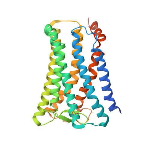
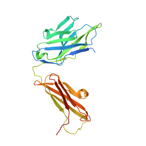
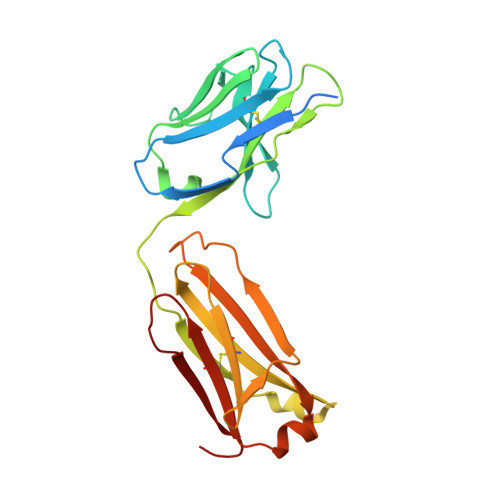
    Prostaglandin E receptor EP4, a G-protein-coupled receptor, is involved in disorders such as cancer and autoimmune disease. Here, we report the crystal structure of human EP4 in complex with its antagonist ONO-AE3-208 and an inhibitory antibody at 3.2 Å resolution. The structure reveals that the extracellular surface is occluded by the extracellular loops and that the antagonist lies at the interface with the lipid bilayer, proximal to the highly conserved Arg316 residue in the seventh transmembrane domain. Functional and docking studies demonstrate that the natural agonist PGE 2 binds in a similar manner. This structural information also provides insight into the ligand entry pathway from the membrane bilayer to the EP4 binding pocket. Furthermore, the structure reveals that the antibody allosterically affects the ligand binding of EP4. These results should facilitate the design of new therapeutic drugs targeting both orthosteric and allosteric sites in this receptor family.


  • Organizational Affiliation

    Department of Medical Chemistry and Cell Biology, Kyoto University Graduate School of Medicine, Konoe-cho, Yoshida, Sakyo-ku, Kyoto, Japan.


Macromolecules
Find similar proteins by:  (by identity cutoff)  |  3D Structure
Entity ID: 1
MoleculeChains Sequence LengthOrganismDetailsImage
Prostaglandin E2 receptor EP4 subtype332Homo sapiensMutation(s): 4 
Gene Names: PTGER4PTGER2
Membrane Entity: Yes 
UniProt & NIH Common Fund Data Resources
Find proteins for P35408 (Homo sapiens)
Explore P35408 
Go to UniProtKB:  P35408
PHAROS:  P35408
GTEx:  ENSG00000171522 
Entity Groups  
Sequence Clusters30% Identity50% Identity70% Identity90% Identity95% Identity100% Identity
UniProt GroupP35408
Sequence Annotations
Expand
  • Reference Sequence
Find similar proteins by:  (by identity cutoff)  |  3D Structure
Entity ID: 2
MoleculeChains Sequence LengthOrganismDetailsImage
Heavy chain of Fab fragmentB [auth H]253Mus musculusMutation(s): 0 
Entity Groups  
Sequence Clusters30% Identity50% Identity70% Identity90% Identity95% Identity100% Identity
Sequence Annotations
Expand
  • Reference Sequence
Find similar proteins by:  (by identity cutoff)  |  3D Structure
Entity ID: 3
MoleculeChains Sequence LengthOrganismDetailsImage
Light chain of Fab fragmentC [auth L]236Mus musculusMutation(s): 0 
Entity Groups  
Sequence Clusters30% Identity50% Identity70% Identity90% Identity95% Identity100% Identity
Sequence Annotations
Expand
  • Reference Sequence
Small Molecules
Ligands 1 Unique
IDChains Name / Formula / InChI Key2D Diagram3D Interactions
8VL
Query on 8VL

Download Ideal Coordinates CCD File 
D [auth A]4-[2-[[(2R)-2-(4-bromanylnaphthalen-1-yl)propanoyl]amino]-4-cyano-phenyl]butanoic acid
C24 H21 Br N2 O3
MCJIJRUBPRBSIT-OAHLLOKOSA-N
Experimental Data & Validation

Experimental Data

Unit Cell:
Length ( Å )Angle ( ˚ )
a = 101.97α = 90
b = 392.23β = 90
c = 81.08γ = 90
Software Package:
Software NamePurpose
XSCALEdata scaling
PHENIXrefinement
PDB_EXTRACTdata extraction
XDSdata reduction
PHENIXphasing

Structure Validation

View Full Validation Report


Ligand Structure Quality Assessment 


Entry History & Funding Information

Deposition Data


Funding OrganizationLocationGrant Number
Japan Science and Technology AgencyJapan--
Japan Agency for Medical Research and DevelopmentJapan--

Revision History  (Full details and data files)

  • Version 1.0: 2018-12-05
    Type: Initial release
  • Version 1.1: 2018-12-12
    Changes: Data collection, Structure summary
  • Version 1.2: 2018-12-19
    Changes: Data collection, Database references
  • Version 1.3: 2019-01-16
    Changes: Data collection, Structure summary
  • Version 1.4: 2019-01-30
    Changes: Data collection, Structure summary
  • Version 1.5: 2023-11-22
    Changes: Data collection, Database references, Refinement description