5XGN

Crystal structure of EGFR 696-1022 T790M/C797S in complex with Go6976


Experimental Data Snapshot

  • Method: X-RAY DIFFRACTION
  • Resolution: 3.00 Å
  • R-Value Free: 0.251 
  • R-Value Work: 0.221 
  • R-Value Observed: 0.222 

wwPDB Validation   3D Report Full Report


Ligand Structure Quality Assessment 


This is version 1.1 of the entry. See complete history


Literature

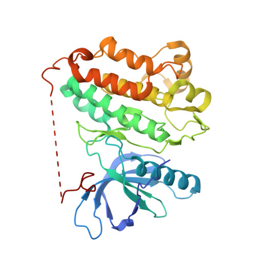
Structural pharmacological studies on EGFR T790M/C797S.

Kong, L.L.Ma, R.Yao, M.Y.Yan, X.E.Zhu, S.J.Zhao, P.Yun, C.H.

(2017) Biochem Biophys Res Commun 488: 266-272

  • DOI: https://doi.org/10.1016/j.bbrc.2017.04.138
  • Primary Citation of Related Structures:  
    5XGM, 5XGN

  • PubMed Abstract: 

    Drug-resistance is a major challenge in targeted therapy of EGFR mutated non-small cell lung cancers (NSCLCs). The third-generation irreversible inhibitors such as AZD9291, CO-1686 and WZ4002 can overcome EGFR T790M drug-resistance mutant through covalent binding through Cys 797, but ultimately lose their efficacy upon emergence of the new mutation C797S. To develop new reversible inhibitors not relying on covalent binding through Cys 797 is therefore urgently demanded. Gö6976 is a staurosporine-like reversible inhibitor targeting T790M while sparing the wild-type EGFR. In the present work, we reported the complex crystal structures of EGFR T790M/C797S + Gö6976 and T790M + Gö6976, along with enzyme kinetic data of EGFR wild-type, T790M and T790M/C797S. These data showed that the C797S mutation does not significantly alter the structure and function of the EGFR kinase, but increases the local hydrophilicity around residue 797. The complex crystal structures also elucidated the detailed binding mode of Gö6976 to EGFR and explained why this compound prefers binding to T790M mutant. These structural pharmacological data would facilitate future drug development studies.


  • Organizational Affiliation

    Institute of Systems Biomedicine, School of Basic Medical Sciences, Peking University Health Science Center, Beijing 100191, China; Department of Biophysics, School of Basic Medical Sciences, Peking University Health Science Center, Beijing 100191, China; Beijing Key Laboratory of Tumor Systems Biology, School of Basic Medical Sciences, Peking University Health Science Center, Beijing 100191, China.


Macromolecules
Find similar proteins by:  (by identity cutoff)  |  3D Structure
Entity ID: 1
MoleculeChains Sequence LengthOrganismDetailsImage
Epidermal growth factor receptor
A, B
331Homo sapiensMutation(s): 2 
Gene Names: EGFRERBBERBB1HER1
EC: 2.7.10.1
UniProt & NIH Common Fund Data Resources
Find proteins for P00533 (Homo sapiens)
Explore P00533 
Go to UniProtKB:  P00533
PHAROS:  P00533
GTEx:  ENSG00000146648 
Entity Groups  
Sequence Clusters30% Identity50% Identity70% Identity90% Identity95% Identity100% Identity
UniProt GroupP00533
Sequence Annotations
Expand
  • Reference Sequence
Small Molecules
Ligands 2 Unique
IDChains Name / Formula / InChI Key2D Diagram3D Interactions
85X
Query on 85X

Download Ideal Coordinates CCD File 
C [auth A]12-(2-Cyanoethyl)-6,7,12,13-tetrahydro-13-methyl-5-oxo-5H-indolo[2,3-a]pyrrolo[3,4-c]carbazole
C24 H18 N4 O
VWVYILCFSYNJHF-UHFFFAOYSA-N
CL
Query on CL

Download Ideal Coordinates CCD File 
D [auth B],
E [auth B]
CHLORIDE ION
Cl
VEXZGXHMUGYJMC-UHFFFAOYSA-M
Experimental Data & Validation

Experimental Data

  • Method: X-RAY DIFFRACTION
  • Resolution: 3.00 Å
  • R-Value Free: 0.251 
  • R-Value Work: 0.221 
  • R-Value Observed: 0.222 
  • Space Group: P 21 21 21
Unit Cell:
Length ( Å )Angle ( ˚ )
a = 49.434α = 90
b = 90.509β = 90
c = 164.223γ = 90
Software Package:
Software NamePurpose
PHENIXrefinement
DENZOdata reduction
HKL-3000data scaling
PHASERphasing

Structure Validation

View Full Validation Report


Ligand Structure Quality Assessment 


Entry History 

Deposition Data

Revision History  (Full details and data files)

  • Version 1.0: 2017-10-11
    Type: Initial release
  • Version 1.1: 2023-11-22
    Changes: Data collection, Database references, Refinement description