5XBM

Structure of SCARB2-JL2 complex


Experimental Data Snapshot

  • Method: X-RAY DIFFRACTION
  • Resolution: 3.50 Å
  • R-Value Free: 0.293 
  • R-Value Work: 0.238 
  • R-Value Observed: 0.241 

wwPDB Validation   3D Report Full Report


Ligand Structure Quality Assessment 


This is version 2.1 of the entry. See complete history


Literature

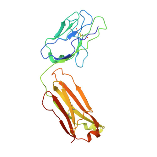
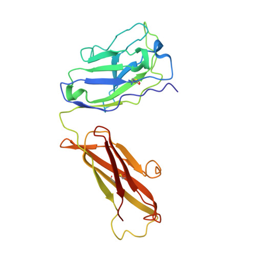
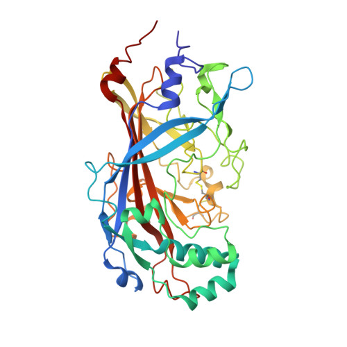
The binding of a monoclonal antibody to the apical region of SCARB2 blocks EV71 infection.

Zhang, X.Yang, P.Wang, N.Zhang, J.Li, J.Guo, H.Yin, X.Rao, Z.Wang, X.Zhang, L.

(2017) Protein Cell 8: 590-600

  • DOI: https://doi.org/10.1007/s13238-017-0405-7
  • Primary Citation of Related Structures:  
    5XBM

  • PubMed Abstract: 

    Entero virus 71 (EV71) causes hand, foot, and mouth disease (HFMD) and occasionally leads to severe neurological complications and even death. Scavenger receptor class B member 2 (SCARB2) is a functional receptor for EV71, that mediates viral attachment, internalization, and uncoating. However, the exact binding site of EV71 on SCARB2 is unknown. In this study, we generated a monoclonal antibody (mAb) that binds to human but not mouse SCARB2. It is named JL2, and it can effectively inhibit EV71 infection of target cells. Using a set of chimeras of human and mouse SCARB2, we identified that the region containing residues 77-113 of human SCARB2 contributes significantly to JL2 binding. The structure of the SCARB2-JL2 complex revealed that JL2 binds to the apical region of SCARB2 involving α-helices 2, 5, and 14. Our results provide new insights into the potential binding sites for EV71 on SCARB2 and the molecular mechanism of EV71 entry.


  • Organizational Affiliation

    Key Laboratory of Infection and Immunity, Institute of Biophysics, Chinese Academy of Sciences, Beijing, 100101, China.


Macromolecules
Find similar proteins by:  (by identity cutoff)  |  3D Structure
Entity ID: 1
MoleculeChains Sequence LengthOrganismDetailsImage
light chain of JL2
A, D
213Mus musculusMutation(s): 0 
Entity Groups  
Sequence Clusters30% Identity50% Identity70% Identity90% Identity95% Identity100% Identity
Sequence Annotations
Expand
  • Reference Sequence
Find similar proteins by:  (by identity cutoff)  |  3D Structure
Entity ID: 2
MoleculeChains Sequence LengthOrganismDetailsImage
heavy chain of JL2
B, E
222Mus musculusMutation(s): 0 
Entity Groups  
Sequence Clusters30% Identity50% Identity70% Identity90% Identity95% Identity100% Identity
Sequence Annotations
Expand
  • Reference Sequence
Find similar proteins by:  (by identity cutoff)  |  3D Structure
Entity ID: 3
MoleculeChains Sequence LengthOrganismDetailsImage
Lysosome membrane protein 2
C, F
394Homo sapiensMutation(s): 0 
Gene Names: SCARB2CD36L2LIMP2LIMPII
UniProt & NIH Common Fund Data Resources
Find proteins for Q14108 (Homo sapiens)
Explore Q14108 
Go to UniProtKB:  Q14108
PHAROS:  Q14108
GTEx:  ENSG00000138760 
Entity Groups  
Sequence Clusters30% Identity50% Identity70% Identity90% Identity95% Identity100% Identity
UniProt GroupQ14108
Sequence Annotations
Expand
  • Reference Sequence
Oligosaccharides

Help

Entity ID: 4
MoleculeChains Length2D Diagram Glycosylation3D Interactions
alpha-D-mannopyranose-(1-3)-beta-D-mannopyranose-(1-4)-2-acetamido-2-deoxy-beta-D-glucopyranose-(1-4)-2-acetamido-2-deoxy-beta-D-glucopyranose
G, M, R
4N-Glycosylation
Glycosylation Resources
GlyTouCan:  G81315DD
GlyCosmos:  G81315DD
GlyGen:  G81315DD
Entity ID: 5
MoleculeChains Length2D Diagram Glycosylation3D Interactions
2-acetamido-2-deoxy-beta-D-glucopyranose-(1-4)-2-acetamido-2-deoxy-beta-D-glucopyranose
H, I, J, N, O
H, I, J, N, O, P
2N-Glycosylation
Glycosylation Resources
GlyTouCan:  G42666HT
GlyCosmos:  G42666HT
GlyGen:  G42666HT
Entity ID: 6
MoleculeChains Length2D Diagram Glycosylation3D Interactions
alpha-D-mannopyranose-(1-3)-[alpha-D-mannopyranose-(1-6)]beta-D-mannopyranose-(1-4)-2-acetamido-2-deoxy-beta-D-glucopyranose-(1-4)-2-acetamido-2-deoxy-beta-D-glucopyranose
K, L, Q
5N-Glycosylation
Glycosylation Resources
GlyTouCan:  G22768VO
GlyCosmos:  G22768VO
GlyGen:  G22768VO
Small Molecules
Ligands 1 Unique
IDChains Name / Formula / InChI Key2D Diagram3D Interactions
NAG
Query on NAG

Download Ideal Coordinates CCD File 
S [auth C],
T [auth F]
2-acetamido-2-deoxy-beta-D-glucopyranose
C8 H15 N O6
OVRNDRQMDRJTHS-FMDGEEDCSA-N
Experimental Data & Validation

Experimental Data

  • Method: X-RAY DIFFRACTION
  • Resolution: 3.50 Å
  • R-Value Free: 0.293 
  • R-Value Work: 0.238 
  • R-Value Observed: 0.241 
  • Space Group: C 1 2 1
Unit Cell:
Length ( Å )Angle ( ˚ )
a = 199.863α = 90
b = 75.646β = 100.11
c = 164.305γ = 90
Software Package:
Software NamePurpose
PHENIXrefinement
HKL-2000data scaling
HKL-2000data reduction
PHENIXphasing

Structure Validation

View Full Validation Report


Ligand Structure Quality Assessment 


Entry History 

Deposition Data

Revision History  (Full details and data files)

  • Version 1.0: 2018-06-27
    Type: Initial release
  • Version 2.0: 2020-07-29
    Type: Remediation
    Reason: Carbohydrate remediation
    Changes: Advisory, Atomic model, Data collection, Derived calculations, Structure summary
  • Version 2.1: 2023-11-22
    Changes: Data collection, Database references, Derived calculations, Refinement description, Structure summary