5X4M

Crystal structure of the BCL6 BTB domain in complex with Compound 1


Experimental Data Snapshot

  • Method: X-RAY DIFFRACTION
  • Resolution: 1.65 Å
  • R-Value Free: 0.206 
  • R-Value Work: 0.179 
  • R-Value Observed: 0.180 

wwPDB Validation   3D Report Full Report


Ligand Structure Quality Assessment 


This is version 1.2 of the entry. See complete history


Literature

Discovery of a B-Cell Lymphoma 6 Protein-Protein Interaction Inhibitor by a Biophysics-Driven Fragment-Based Approach

Kamada, Y.Sakai, N.Sogabe, S.Ida, K.Oki, H.Sakamoto, K.Lane, W.Snell, G.Iida, M.Imaeda, Y.Sakamoto, J.Matsui, J.

(2017) J Med Chem 60: 4358-4368

  • DOI: https://doi.org/10.1021/acs.jmedchem.7b00313
  • Primary Citation of Related Structures:  
    5X4M, 5X4N, 5X4O, 5X4P, 5X4Q

  • PubMed Abstract: 

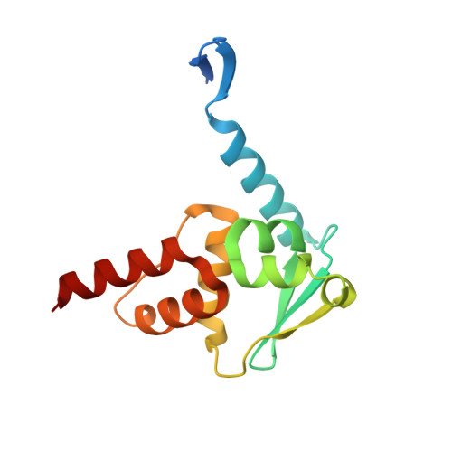
    B-cell lymphoma 6 (BCL6) is a transcriptional factor that expresses in lymphocytes and regulates the differentiation and proliferation of lymphocytes. Therefore, BCL6 is a therapeutic target for autoimmune diseases and cancer treatment. This report presents the discovery of BCL6-corepressor interaction inhibitors by using a biophysics-driven fragment-based approach. Using the surface plasmon resonance (SPR)-based fragment screening, we successfully identified fragment 1 (SPR K D = 1200 μM, ligand efficiency (LE) = 0.28), a competitive binder to the natural ligand BCoR peptide. Moreover, we elaborated 1 into the more potent compound 7 (SPR K D = 0.078 μM, LE = 0.37, cell-free protein-protein interaction (PPI) IC 50 = 0.48 μM (ELISA), cellular PPI IC 50 = 8.6 μM (M2H)) by a structure-based design and structural integration with a second high-throughput screening hit.


  • Organizational Affiliation

    Takeda Pharmaceutical Company Limited , 26-1 Muraoka-Higashi 2-chome, Fujisawa, Kanagawa 251-8555, Japan.


Macromolecules
Find similar proteins by:  (by identity cutoff)  |  3D Structure
Entity ID: 1
MoleculeChains Sequence LengthOrganismDetailsImage
B-cell lymphoma 6 protein141Homo sapiensMutation(s): 0 
Gene Names: BCL6BCL5LAZ3ZBTB27ZNF51
UniProt & NIH Common Fund Data Resources
Find proteins for P41182 (Homo sapiens)
Explore P41182 
Go to UniProtKB:  P41182
PHAROS:  P41182
GTEx:  ENSG00000113916 
Entity Groups  
Sequence Clusters30% Identity50% Identity70% Identity90% Identity95% Identity100% Identity
UniProt GroupP41182
Sequence Annotations
Expand
  • Reference Sequence
Small Molecules
Ligands 1 Unique
IDChains Name / Formula / InChI Key2D Diagram3D Interactions
7ZF
Query on 7ZF

Download Ideal Coordinates CCD File 
B [auth A]N-phenyl-1,3,5-triazine-2,4-diamine
C9 H9 N5
YMFGJWGABDOFID-UHFFFAOYSA-N
Binding Affinity Annotations 
IDSourceBinding Affinity
7ZF BindingDB:  5X4M Kd: 1.20e+6 (nM) from 1 assay(s)
Binding MOAD:  5X4M Kd: 1.20e+6 (nM) from 1 assay(s)
Experimental Data & Validation

Experimental Data

  • Method: X-RAY DIFFRACTION
  • Resolution: 1.65 Å
  • R-Value Free: 0.206 
  • R-Value Work: 0.179 
  • R-Value Observed: 0.180 
  • Space Group: P 61 2 2
Unit Cell:
Length ( Å )Angle ( ˚ )
a = 66.425α = 90
b = 66.425β = 90
c = 152.192γ = 120
Software Package:
Software NamePurpose
REFMACrefinement
HKL-2000data reduction
SCALEPACKdata scaling
MOLREPphasing

Structure Validation

View Full Validation Report


Ligand Structure Quality Assessment 


Entry History 

Deposition Data

Revision History  (Full details and data files)

  • Version 1.0: 2017-05-24
    Type: Initial release
  • Version 1.1: 2017-06-07
    Changes: Database references
  • Version 1.2: 2023-11-22
    Changes: Data collection, Database references, Refinement description