5VAR

Crystal structure of KDM4A tandem TUDOR domain in complex with a tri-methyl lysine competitive inhibitor


Experimental Data Snapshot

  • Method: X-RAY DIFFRACTION
  • Resolution: 1.83 Å
  • R-Value Free: 0.243 
  • R-Value Work: 0.194 
  • R-Value Observed: 0.196 

wwPDB Validation   3D Report Full Report


Ligand Structure Quality Assessment 


This is version 1.4 of the entry. See complete history


Literature

Targeting lysine specific demethylase 4A (KDM4A) tandem TUDOR domain - A fragment based approach.

Upadhyay, A.K.Judge, R.A.Li, L.Pithawalla, R.Simanis, J.Bodelle, P.M.Marin, V.L.Henry, R.F.Petros, A.M.Sun, C.

(2018) Bioorg Med Chem Lett 28: 1708-1713

  • DOI: https://doi.org/10.1016/j.bmcl.2018.04.050
  • Primary Citation of Related Structures:  
    5VAR

  • PubMed Abstract: 

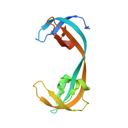
    The tandem TUDOR domains present in the non-catalytic C-terminal half of the KDM4A, 4B and 4C enzymes play important roles in regulating their chromatin localizations and substrate specificities. They achieve this regulatory role by binding to different tri-methylated lysine residues on histone H3 (H3-K4me3, H3-K23me3) and histone H4 (H4-K20me3) depending upon the specific chromatin environment. In this work, we have used a 2D-NMR based fragment screening approach to identify a novel fragment (1a), which binds to the KDM4A-TUDOR domain and shows modest competition with H3-K4me3 binding in biochemical as well as in vitro cell based assays. A co-crystal structure of KDM4A TUDOR domain in complex with 1a shows that the fragment binds stereo-specifically to the methyl lysine binding pocket forming a network of strong hydrogen bonds and hydrophobic interactions. We anticipate that the fragment 1a can be further developed into a novel allosteric inhibitor of the KDM4 family of enzymes through targeting their C-terminal tandem TUDOR domain.


  • Organizational Affiliation

    AbbVie Inc., 1 North Waukegan Road, North Chicago, IL 60064, USA. Electronic address: anup.upadhyay@abbvie.com.


Macromolecules
Find similar proteins by:  (by identity cutoff)  |  3D Structure
Entity ID: 1
MoleculeChains Sequence LengthOrganismDetailsImage
Lysine-specific demethylase 4A119Homo sapiensMutation(s): 0 
Gene Names: KDM4AJHDM3AJMJD2JMJD2AKIAA0677
EC: 1.14.11
UniProt & NIH Common Fund Data Resources
Find proteins for O75164 (Homo sapiens)
Explore O75164 
Go to UniProtKB:  O75164
PHAROS:  O75164
GTEx:  ENSG00000066135 
Entity Groups  
Sequence Clusters30% Identity50% Identity70% Identity90% Identity95% Identity100% Identity
UniProt GroupO75164
Sequence Annotations
Expand
  • Reference Sequence
Small Molecules
Ligands 1 Unique
IDChains Name / Formula / InChI Key2D Diagram3D Interactions
92Y
Query on 92Y

Download Ideal Coordinates CCD File 
B [auth A](1R,2S,3R,4S)-3-[(dimethylamino)methyl]-1-phenylbicyclo[2.2.1]heptan-2-ol
C16 H23 N O
HGNFUCJMHJYHIN-QCEMKRCNSA-N
Binding Affinity Annotations 
IDSourceBinding Affinity
92Y BindingDB:  5VAR Kd: 8.00e+4 (nM) from 1 assay(s)
Binding MOAD:  5VAR Kd: 8.17e+4 (nM) from 1 assay(s)
Experimental Data & Validation

Experimental Data

  • Method: X-RAY DIFFRACTION
  • Resolution: 1.83 Å
  • R-Value Free: 0.243 
  • R-Value Work: 0.194 
  • R-Value Observed: 0.196 
  • Space Group: P 1 21 1
Unit Cell:
Length ( Å )Angle ( ˚ )
a = 33.27α = 90
b = 39.84β = 101.61
c = 44.3γ = 90
Software Package:
Software NamePurpose
Aimlessdata scaling
MOLREPphasing
BUSTERrefinement
PDB_EXTRACTdata extraction
XDSdata reduction

Structure Validation

View Full Validation Report


Ligand Structure Quality Assessment 


Entry History 

Deposition Data

Revision History  (Full details and data files)

  • Version 1.0: 2018-03-28
    Type: Initial release
  • Version 1.1: 2018-05-09
    Changes: Data collection, Database references
  • Version 1.2: 2018-05-16
    Changes: Data collection, Database references
  • Version 1.3: 2018-09-05
    Changes: Data collection, Database references
  • Version 1.4: 2023-10-04
    Changes: Data collection, Database references, Refinement description