5MG7

New Insights into the Role of DNA Shape on Its Recognition by p53 Proteins (complex p53DBD-p53R2)


Experimental Data Snapshot

  • Method: X-RAY DIFFRACTION
  • Resolution: 1.45 Å
  • R-Value Free: 0.202 
  • R-Value Work: 0.171 
  • R-Value Observed: 0.172 

wwPDB Validation   3D Report Full Report


This is version 1.4 of the entry. See complete history


Literature

New Insights into the Role of DNA Shape on Its Recognition by p53 Proteins.

Golovenko, D.Brauning, B.Vyas, P.Haran, T.E.Rozenberg, H.Shakked, Z.

(2018) Structure 26: 1237-1250.e6

  • DOI: https://doi.org/10.1016/j.str.2018.06.006
  • Primary Citation of Related Structures:  
    5MCT, 5MCU, 5MCV, 5MCW, 5MF7, 5MG7, 6FJ5

  • PubMed Abstract: 

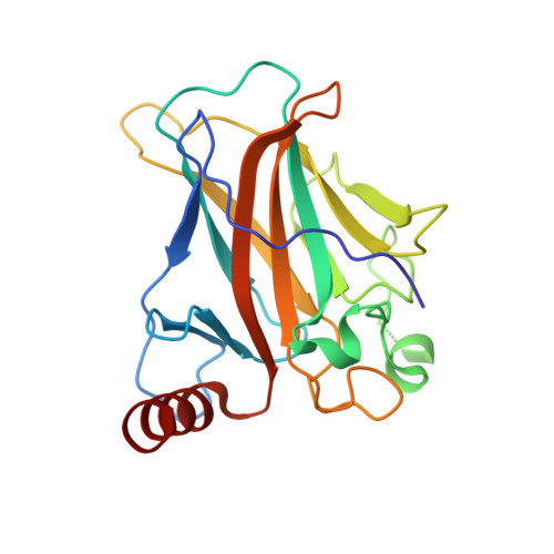
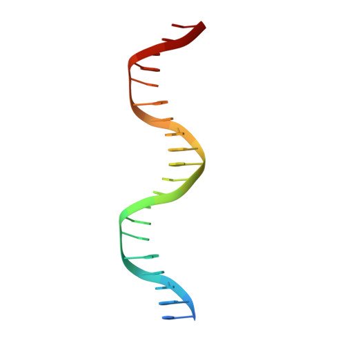
    The tumor suppressor p53 acts as a transcription factor recognizing diverse DNA response elements (REs). Previous structural studies of p53-DNA complexes revealed non-canonical Hoogsteen geometry of A/T base pairs at conserved CATG motifs leading to changes in DNA shape and its interface with p53. To study the effects of DNA shape on binding characteristics, we designed REs with modified base pairs "locked" into either Hoogsteen or Watson-Crick form. Here we present crystal structures of these complexes and their thermodynamic and kinetic parameters, demonstrating that complexes with Hoogsteen base pairs are stabilized relative to those with all-Watson-Crick base pairs. CATG motifs are abundant in p53REs such as GADD45 and p53R2 related to cell-cycle arrest and DNA repair. The high-resolution structures of these complexes validate their propensity to adopt the unique Hoogsteen-induced structure, thus providing insights into the functional role of DNA shape and broadening the mechanisms that contribute to DNA recognition by proteins.


  • Organizational Affiliation

    Department of Structural Biology, Weizmann Institute of Science, Rehovot 76100, Israel.


Macromolecules

Find similar proteins by:  (by identity cutoff)  |  3D Structure
Entity ID: 1
MoleculeChains Sequence LengthOrganismDetailsImage
Cellular tumor antigen p53
A, B
200Homo sapiensMutation(s): 0 
Gene Names: TP53P53
UniProt & NIH Common Fund Data Resources
Find proteins for P04637 (Homo sapiens)
Explore P04637 
Go to UniProtKB:  P04637
PHAROS:  P04637
GTEx:  ENSG00000141510 
Entity Groups  
Sequence Clusters30% Identity50% Identity70% Identity90% Identity95% Identity100% Identity
UniProt GroupP04637
Sequence Annotations
Expand
  • Reference Sequence

Find similar nucleic acids by:  Sequence   |   3D Structure  

Entity ID: 2
MoleculeChains LengthOrganismImage
DNA21synthetic construct
Sequence Annotations
Expand
  • Reference Sequence
Experimental Data & Validation

Experimental Data

  • Method: X-RAY DIFFRACTION
  • Resolution: 1.45 Å
  • R-Value Free: 0.202 
  • R-Value Work: 0.171 
  • R-Value Observed: 0.172 
  • Space Group: C 1 2 1
Unit Cell:
Length ( Å )Angle ( ˚ )
a = 137.621α = 90
b = 49.804β = 93.34
c = 68.089γ = 90
Software Package:
Software NamePurpose
PHENIXrefinement
PHASERphasing
HKL-2000data collection
HKL-2000data scaling
PDB_EXTRACTdata extraction
HKL-2000data reduction

Structure Validation

View Full Validation Report


Entry History 

Deposition Data

Revision History  (Full details and data files)

  • Version 1.0: 2018-06-13
    Type: Initial release
  • Version 1.1: 2018-06-27
    Changes: Data collection, Database references
  • Version 1.2: 2019-01-02
    Changes: Data collection, Database references, Structure summary
  • Version 1.3: 2019-10-30
    Changes: Data collection, Derived calculations
  • Version 1.4: 2024-01-17
    Changes: Data collection, Database references, Refinement description