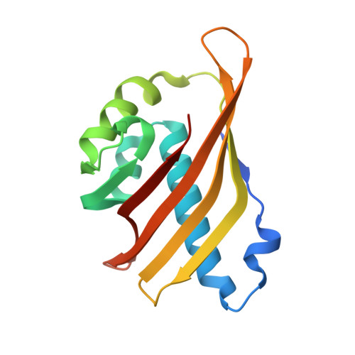
5IG3

Crystal structure of the human CaMKII-alpha hub


Experimental Data Snapshot

  • Method: X-RAY DIFFRACTION
  • Resolution: 2.75 Å
  • R-Value Free: 0.268 
  • R-Value Work: 0.220 
  • R-Value Observed: 0.223 

wwPDB Validation   3D Report Full Report


This is version 1.5 of the entry. See complete history


Literature

Molecular mechanism of activation-triggered subunit exchange in Ca(2+)/calmodulin-dependent protein kinase II.

Bhattacharyya, M.Stratton, M.M.Going, C.C.McSpadden, E.D.Huang, Y.Susa, A.C.Elleman, A.Cao, Y.M.Pappireddi, N.Burkhardt, P.Gee, C.L.Barros, T.Schulman, H.Williams, E.R.Kuriyan, J.

(2016) Elife 5

  • DOI: https://doi.org/10.7554/eLife.13405
  • Primary Citation of Related Structures:  
    5IG0, 5IG1, 5IG3, 5IG4, 5IG5

  • PubMed Abstract: 

    Activation triggers the exchange of subunits in Ca(2+)/calmodulin-dependent protein kinase II (CaMKII), an oligomeric enzyme that is critical for learning, memory, and cardiac function. The mechanism by which subunit exchange occurs remains elusive. We show that the human CaMKII holoenzyme exists in dodecameric and tetradecameric forms, and that the calmodulin (CaM)-binding element of CaMKII can bind to the hub of the holoenzyme and destabilize it to release dimers. The structures of CaMKII from two distantly diverged organisms suggest that the CaM-binding element of activated CaMKII acts as a wedge by docking at intersubunit interfaces in the hub. This converts the hub into a spiral form that can release or gain CaMKII dimers. Our data reveal a three-way competition for the CaM-binding element, whereby phosphorylation biases it towards the hub interface, away from the kinase domain and calmodulin, thus unlocking the ability of activated CaMKII holoenzymes to exchange dimers with unactivated ones.


  • Organizational Affiliation

    Department of Molecular and Cell Biology, University of California, Berkeley, Berkeley, United States.


Macromolecules
Find similar proteins by:  (by identity cutoff)  |  3D Structure
Entity ID: 1
MoleculeChains Sequence LengthOrganismDetailsImage
Calcium/calmodulin-dependent protein kinase type II subunit alpha
A, B, C, D, E
A, B, C, D, E, F
153Homo sapiensMutation(s): 0 
Gene Names: CAMK2ACAMKAKIAA0968
EC: 2.7.11.17
UniProt & NIH Common Fund Data Resources
Find proteins for Q9UQM7 (Homo sapiens)
Explore Q9UQM7 
Go to UniProtKB:  Q9UQM7
PHAROS:  Q9UQM7
GTEx:  ENSG00000070808 
Entity Groups  
Sequence Clusters30% Identity50% Identity70% Identity90% Identity95% Identity100% Identity
UniProt GroupQ9UQM7
Sequence Annotations
Expand
  • Reference Sequence
Experimental Data & Validation

Experimental Data

  • Method: X-RAY DIFFRACTION
  • Resolution: 2.75 Å
  • R-Value Free: 0.268 
  • R-Value Work: 0.220 
  • R-Value Observed: 0.223 
  • Space Group: P 43 21 2
Unit Cell:
Length ( Å )Angle ( ˚ )
a = 89.888α = 90
b = 89.888β = 90
c = 226.577γ = 90
Software Package:
Software NamePurpose
PHENIXrefinement
XDSdata reduction
Aimlessdata scaling
PHASERphasing

Structure Validation

View Full Validation Report


Entry History & Funding Information

Deposition Data


Funding OrganizationLocationGrant Number
Howard Hughes Medical Institute (HHMI)United StatesNA

Revision History  (Full details and data files)

  • Version 1.0: 2016-03-23
    Type: Initial release
  • Version 1.1: 2016-04-06
    Changes: Structure summary
  • Version 1.2: 2016-07-13
    Changes: Data collection
  • Version 1.3: 2017-09-27
    Changes: Author supporting evidence, Derived calculations
  • Version 1.4: 2019-11-20
    Changes: Author supporting evidence
  • Version 1.5: 2023-09-27
    Changes: Data collection, Database references, Refinement description