5HPT

System-wide modulation of HECT E3 ligases with selective ubiquitin variant probes: WWP1, Ubv P2.3 and UBCH7


Experimental Data Snapshot

  • Method: X-RAY DIFFRACTION
  • Resolution: 2.84 Å
  • R-Value Free: 0.237 
  • R-Value Work: 0.218 
  • R-Value Observed: 0.219 

wwPDB Validation   3D Report Full Report


This is version 1.6 of the entry. See complete history


Literature

System-Wide Modulation of HECT E3 Ligases with Selective Ubiquitin Variant Probes.

Zhang, W.Wu, K.P.Sartori, M.A.Kamadurai, H.B.Ordureau, A.Jiang, C.Mercredi, P.Y.Murchie, R.Hu, J.Persaud, A.Mukherjee, M.Li, N.Doye, A.Walker, J.R.Sheng, Y.Hao, Z.Li, Y.Brown, K.R.Lemichez, E.Chen, J.Tong, Y.Harper, J.W.Moffat, J.Rotin, D.Schulman, B.A.Sidhu, S.S.

(2016) Mol Cell 62: 121-136

  • DOI: https://doi.org/10.1016/j.molcel.2016.02.005
  • Primary Citation of Related Structures:  
    5C7J, 5C7M, 5HPK, 5HPL, 5HPS, 5HPT

  • PubMed Abstract: 

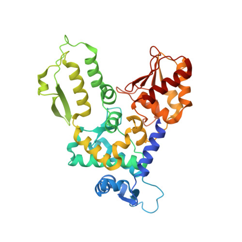
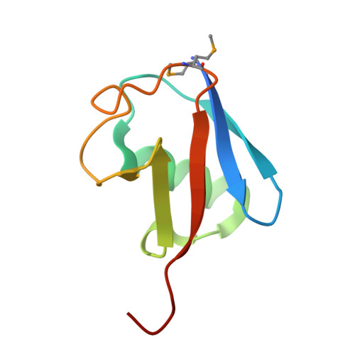
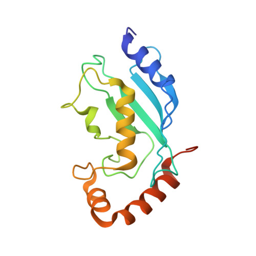
    HECT-family E3 ligases ubiquitinate protein substrates to control virtually every eukaryotic process and are misregulated in numerous diseases. Nonetheless, understanding of HECT E3s is limited by a paucity of selective and potent modulators. To overcome this challenge, we systematically developed ubiquitin variants (UbVs) that inhibit or activate HECT E3s. Structural analysis of 6 HECT-UbV complexes revealed UbV inhibitors hijacking the E2-binding site and activators occupying a ubiquitin-binding exosite. Furthermore, UbVs unearthed distinct regulation mechanisms among NEDD4 subfamily HECTs and proved useful for modulating therapeutically relevant targets of HECT E3s in cells and intestinal organoids, and in a genetic screen that identified a role for NEDD4L in regulating cell migration. Our work demonstrates versatility of UbVs for modulating activity across an E3 family, defines mechanisms and provides a toolkit for probing functions of HECT E3s, and establishes a general strategy for systematic development of modulators targeting families of signaling proteins.


  • Organizational Affiliation

    Donnelly Centre for Cellular and Biomolecular Research, Banting and Best Department of Medical Research, University of Toronto, 160 College Street, Toronto, ON M5S3E1, Canada.


Macromolecules
Find similar proteins by:  (by identity cutoff)  |  3D Structure
Entity ID: 1
MoleculeChains Sequence LengthOrganismDetailsImage
NEDD4-like E3 ubiquitin-protein ligase WWP1
A, D, G
383Homo sapiensMutation(s): 0 
Gene Names: WWP1
EC: 6.3.2
UniProt & NIH Common Fund Data Resources
Find proteins for Q9H0M0 (Homo sapiens)
Explore Q9H0M0 
Go to UniProtKB:  Q9H0M0
PHAROS:  Q9H0M0
GTEx:  ENSG00000123124 
Entity Groups  
Sequence Clusters30% Identity50% Identity70% Identity90% Identity95% Identity100% Identity
UniProt GroupQ9H0M0
Sequence Annotations
Expand
  • Reference Sequence
Find similar proteins by:  (by identity cutoff)  |  3D Structure
Entity ID: 2
MoleculeChains Sequence LengthOrganismDetailsImage
Ubiquitin variant P2.3
B, E, H
83Homo sapiensMutation(s): 0 
UniProt & NIH Common Fund Data Resources
Find proteins for P62987 (Homo sapiens)
Explore P62987 
Go to UniProtKB:  P62987
PHAROS:  P62987
GTEx:  ENSG00000221983 
Entity Groups  
Sequence Clusters30% Identity50% Identity70% Identity90% Identity95% Identity100% Identity
UniProt GroupP62987
Sequence Annotations
Expand
  • Reference Sequence
Find similar proteins by:  (by identity cutoff)  |  3D Structure
Entity ID: 3
MoleculeChains Sequence LengthOrganismDetailsImage
Ubiquitin-conjugating enzyme E2 L3
C, F
161Homo sapiensMutation(s): 0 
Gene Names: UBE2L3UBCE7UBCH7
EC: 2.3.2.23
UniProt & NIH Common Fund Data Resources
Find proteins for P68036 (Homo sapiens)
Explore P68036 
Go to UniProtKB:  P68036
PHAROS:  P68036
GTEx:  ENSG00000185651 
Entity Groups  
Sequence Clusters30% Identity50% Identity70% Identity90% Identity95% Identity100% Identity
UniProt GroupP68036
Sequence Annotations
Expand
  • Reference Sequence
Small Molecules
Modified Residues  1 Unique
IDChains TypeFormula2D DiagramParent
MSE
Query on MSE
B, E, H
L-PEPTIDE LINKINGC5 H11 N O2 SeMET
Experimental Data & Validation

Experimental Data

  • Method: X-RAY DIFFRACTION
  • Resolution: 2.84 Å
  • R-Value Free: 0.237 
  • R-Value Work: 0.218 
  • R-Value Observed: 0.219 
  • Space Group: P 21 21 21
Unit Cell:
Length ( Å )Angle ( ˚ )
a = 114.005α = 90
b = 118.897β = 90
c = 158.663γ = 90
Software Package:
Software NamePurpose
PHENIXrefinement
Cootmodel building
PHASERphasing

Structure Validation

View Full Validation Report


Entry History & Funding Information

Deposition Data


Funding OrganizationLocationGrant Number
National Institutes of Health/National Institute of General Medical Sciences (NIH/NIGMS)United StatesGM103403

Revision History  (Full details and data files)

  • Version 1.0: 2016-03-16
    Type: Initial release
  • Version 1.1: 2016-03-23
    Changes: Database references
  • Version 1.2: 2016-04-20
    Changes: Database references
  • Version 1.3: 2017-09-27
    Changes: Author supporting evidence, Database references, Derived calculations
  • Version 1.4: 2019-12-25
    Changes: Author supporting evidence
  • Version 1.5: 2023-09-27
    Changes: Data collection, Database references, Refinement description
  • Version 1.6: 2023-11-15
    Changes: Data collection