Structural mechanism underlying regulation of human EFhd2/Swiprosin-1 actin-bundling activity by Ser183 phosphorylation.
Park, K.R., An, J.Y., Kang, J.Y., Lee, J.G., Lee, Y., Mun, S.A., Jun, C.D., Song, W.K., Eom, S.H.(2017) Biochem Biophys Res Commun 483: 442-448
- PubMed: 28011271
- DOI: https://doi.org/10.1016/j.bbrc.2016.12.124
- Primary Citation of Related Structures:
5H0P - PubMed Abstract:
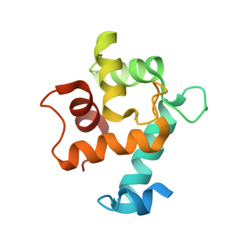
EF-hand domain-containing protein D2/Swiprosin-1 (EFhd2) is an actin-binding protein mainly expressed in the central nervous and the immune systems of mammals. Intracellular events linked to EFhd2, such as membrane protrusion formation, cell adhesion, and BCR signaling, are triggered by the association of EFhd2 and F-actin. We previously reported that Ca 2+ enhances the F-actin-bundling ability of EFhd2 through maintaining a rigid parallel EFhd2-homodimer structure. It was also reported that the F-actin-bundling ability of EFhd2 is regulated by a phosphorylation-dependent mechanism. EGF-induced phosphorylation at Ser183 of EFhd2 has been shown to inhibit F-actin-bundling, leading to irregular actin dynamics at the leading edges of cells. However, the underlying mechanism of this inhibition has remained elusive. Here, we report the crystal structure of a phospho-mimicking mutant (S183E) of the EFhd2 core domain, where the actin-binding sites are located. Although the overall structure of the phospho-mimicking mutant is similar to the one of the unphosphorylated form, we observed a conformational transition from ordered to disordered structure in the linker region at the C-terminus of the mutant. Based on our structural and biochemical analyses, we suggest that phosphorylation at Ser183 of EFhd2 causes changes in the local conformational dynamics and the surface charge distribution of the actin-binding site, resulting in a re-coordination of the actin-binding sites in the dimer structure and a reduction of F-actin-bundling activity without affecting the F-actin-binding capacity.
Organizational Affiliation:
School of Life Science, Gwangju Institute of Science and Technology (GIST), 123 Cheomdangwagi-ro, Buk-gu, Gwangju 61005, Republic of Korea; Steitz Center for Structural Biology, Gwangju Institute of Science and Technology (GIST), 123 Cheomdangwagi-ro, Buk-gu, Gwangju 61005, Republic of Korea.