5E6J

Structure of SARS PLpro bound to a Lys48-linked di-ubiquitin activity based probe


Experimental Data Snapshot

  • Method: X-RAY DIFFRACTION
  • Resolution: 2.85 Å
  • R-Value Free: 0.264 
  • R-Value Work: 0.232 
  • R-Value Observed: 0.234 

wwPDB Validation   3D Report Full Report


This is version 1.7 of the entry. See complete history


Literature

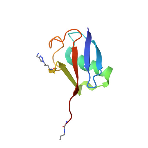
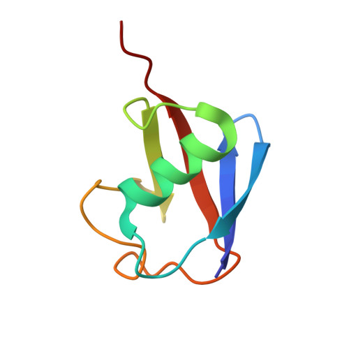
Recognition of Lys48-Linked Di-ubiquitin and Deubiquitinating Activities of the SARS Coronavirus Papain-like Protease.

Bekes, M.van der Heden van Noort, G.J.Ekkebus, R.Ovaa, H.Huang, T.T.Lima, C.D.

(2016) Mol Cell 62: 572-585

  • DOI: https://doi.org/10.1016/j.molcel.2016.04.016
  • Primary Citation of Related Structures:  
    5E6J

  • PubMed Abstract: 

    Deubiquitinating enzymes (DUBs) recognize and cleave linkage-specific polyubiquitin (polyUb) chains, but mechanisms underlying specificity remain elusive in many cases. The severe acute respiratory syndrome (SARS) coronavirus papain-like protease (PLpro) is a DUB that cleaves ISG15, a two-domain Ub-like protein, and Lys48-linked polyUb chains, releasing diUb(Lys48) products. To elucidate this specificity, we report the 2.85 Å crystal structure of SARS PLpro bound to a diUb(Lys48) activity-based probe. SARS PLpro binds diUb(Lys48) in an extended conformation via two contact sites, S1 and S2, which are proximal and distal to the active site, respectively. We show that specificity for polyUb(Lys48) chains is predicated on contacts in the S2 site and enhanced by an S1-S1' preference for a Lys48 linkage across the active site. In contrast, ISG15 specificity is dominated by contacts in the S1 site. Determinants revealed for polyUb(Lys48) specificity should prove useful in understanding PLpro deubiquitinating activities in coronavirus infections.


  • Organizational Affiliation

    Department of Biochemistry and Molecular Pharmacology, New York University School of Medicine, New York, NY 10016, USA; Structural Biology Program, Sloan Kettering Institute, 1275 York Avenue, New York, NY 10065, USA.


Macromolecules
Find similar proteins by:  (by identity cutoff)  |  3D Structure
Entity ID: 1
MoleculeChains Sequence LengthOrganismDetailsImage
Replicase polyprotein 1ab
A, D
325Severe acute respiratory syndrome-related coronavirusMutation(s): 0 
Gene Names: rep1a-1b
EC: 3.4.19.12 (PDB Primary Data), 3.4.22.69 (PDB Primary Data), 3.4.22 (PDB Primary Data), 2.7.7.48 (PDB Primary Data), 3.6.4.12 (PDB Primary Data), 3.6.4.13 (PDB Primary Data), 2.1.1 (PDB Primary Data), 3.1.13 (PDB Primary Data), 3.1 (PDB Primary Data)
UniProt
Find proteins for P0C6X7 (Severe acute respiratory syndrome coronavirus)
Explore P0C6X7 
Go to UniProtKB:  P0C6X7
Entity Groups  
Sequence Clusters30% Identity50% Identity70% Identity90% Identity95% Identity100% Identity
UniProt GroupP0C6X7
Sequence Annotations
Expand
  • Reference Sequence
Find similar proteins by:  (by identity cutoff)  |  3D Structure
Entity ID: 2
MoleculeChains Sequence LengthOrganismDetailsImage
ubiquitin
B, E
76synthetic constructMutation(s): 1 
UniProt & NIH Common Fund Data Resources
Find proteins for P0CG48 (Homo sapiens)
Explore P0CG48 
Go to UniProtKB:  P0CG48
PHAROS:  P0CG48
GTEx:  ENSG00000150991 
Entity Groups  
Sequence Clusters30% Identity50% Identity70% Identity90% Identity95% Identity100% Identity
UniProt GroupP0CG48
Sequence Annotations
Expand
  • Reference Sequence
Find similar proteins by:  (by identity cutoff)  |  3D Structure
Entity ID: 3
MoleculeChains Sequence LengthOrganismDetailsImage
Polyubiquitin-B
C, F
75synthetic constructMutation(s): 0 
UniProt & NIH Common Fund Data Resources
Find proteins for P0CG47 (Homo sapiens)
Explore P0CG47 
Go to UniProtKB:  P0CG47
PHAROS:  P0CG47
GTEx:  ENSG00000170315 
Entity Groups  
Sequence Clusters30% Identity50% Identity70% Identity90% Identity95% Identity100% Identity
UniProt GroupP0CG47
Sequence Annotations
Expand
  • Reference Sequence
Small Molecules
Modified Residues  1 Unique
IDChains TypeFormula2D DiagramParent
5MW
Query on 5MW
B, E
L-PEPTIDE LINKINGC8 H15 N5 O2LYS
Experimental Data & Validation

Experimental Data

  • Method: X-RAY DIFFRACTION
  • Resolution: 2.85 Å
  • R-Value Free: 0.264 
  • R-Value Work: 0.232 
  • R-Value Observed: 0.234 
  • Space Group: P 1 21 1
Unit Cell:
Length ( Å )Angle ( ˚ )
a = 72.981α = 90
b = 68.239β = 103.21
c = 119.021γ = 90
Software Package:
Software NamePurpose
PHENIXrefinement
HKL-2000data reduction
HKL-2000data scaling
PHENIXphasing

Structure Validation

View Full Validation Report


Entry History & Funding Information

Deposition Data


Funding OrganizationLocationGrant Number
National Institutes of Health/National Institute of General Medical Sciences (NIH/NIGMS)United StatesGM065872
National Institutes of Health/National Institute of General Medical Sciences (NIH/NIGMS)United StatesF32GM100598
National Institutes of Health/National Institute of General Medical Sciences (NIH/NIGMS)United StatesGM084244
Howard Hughes Medical Institute (HHMI)United States--
Netherlands Organisation for Scientific Research (NWO)Netherlands724013002

Revision History  (Full details and data files)

  • Version 1.0: 2016-05-18
    Type: Initial release
  • Version 1.1: 2017-01-25
    Changes: Derived calculations
  • Version 1.2: 2017-09-06
    Changes: Author supporting evidence
  • Version 1.3: 2017-11-01
    Changes: Author supporting evidence
  • Version 1.4: 2017-12-20
    Changes: Database references
  • Version 1.5: 2019-11-20
    Changes: Author supporting evidence
  • Version 1.6: 2023-09-27
    Changes: Data collection, Database references, Derived calculations, Refinement description
  • Version 1.7: 2023-11-15
    Changes: Data collection, Derived calculations