5AOK

Structure of the p53 cancer mutant Y220C with bound small molecule PhiKan7099


Experimental Data Snapshot

  • Method: X-RAY DIFFRACTION
  • Resolution: 1.35 Å
  • R-Value Free: 0.169 
  • R-Value Work: 0.145 
  • R-Value Observed: 0.146 

wwPDB Validation   3D Report Full Report


Ligand Structure Quality Assessment 


This is version 1.3 of the entry. See complete history


Literature

Exploiting Transient Protein States for the Design of Small-Molecule Stabilizers of Mutant P53.

Joerger, A.C.Bauer, M.R.Wilcken, R.Baud, M.G.J.Harbrecht, H.Exner, T.E.Boeckler, F.M.Spencer, J.Fersht, A.R.

(2015) Structure 23: 2246

  • DOI: https://doi.org/10.1016/j.str.2015.10.016
  • Primary Citation of Related Structures:  
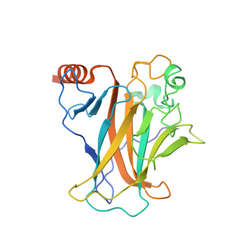
    5AB9, 5ABA, 5AOI, 5AOJ, 5AOK, 5AOL, 5AOM

  • PubMed Abstract: 

    The destabilizing p53 cancer mutation Y220C creates an extended crevice on the surface of the protein that can be targeted by small-molecule stabilizers. Here, we identify different classes of small molecules that bind to this crevice and determine their binding modes by X-ray crystallography. These structures reveal two major conformational states of the pocket and a cryptic, transiently open hydrophobic subpocket that is modulated by Cys220. In one instance, specifically targeting this transient protein state by a pyrrole moiety resulted in a 40-fold increase in binding affinity. Molecular dynamics simulations showed that both open and closed states of this subsite were populated at comparable frequencies along the trajectories. Our data extend the framework for the design of high-affinity Y220C mutant binders for use in personalized anticancer therapy and, more generally, highlight the importance of implementing protein dynamics and hydration patterns in the drug-discovery process.


  • Organizational Affiliation

    MRC Laboratory of Molecular Biology, Francis Crick Avenue, Cambridge CB2 0QH, UK. Electronic address: andreas.joerger@gmx.de.


Macromolecules
Find similar proteins by:  (by identity cutoff)  |  3D Structure
Entity ID: 1
MoleculeChains Sequence LengthOrganismDetailsImage
CELLULAR TUMOR ANTIGEN P53
A, B
219Homo sapiensMutation(s): 5 
UniProt & NIH Common Fund Data Resources
Find proteins for P04637 (Homo sapiens)
Explore P04637 
Go to UniProtKB:  P04637
PHAROS:  P04637
GTEx:  ENSG00000141510 
Entity Groups  
Sequence Clusters30% Identity50% Identity70% Identity90% Identity95% Identity100% Identity
UniProt GroupP04637
Sequence Annotations
Expand
  • Reference Sequence
Small Molecules
Ligands 4 Unique
IDChains Name / Formula / InChI Key2D Diagram3D Interactions
GOH
Query on GOH

Download Ideal Coordinates CCD File 
E [auth A],
I [auth B]
5-[2-cyclopropyl-5-(1H-pyrrol-1-yl)-1,3-oxazol-4-yl]-1H-1,2,3,4-tetrazole
C11 H10 N6 O
WNHQAZRUEGMEKF-UHFFFAOYSA-N
PEG
Query on PEG

Download Ideal Coordinates CCD File 
H [auth B]DI(HYDROXYETHYL)ETHER
C4 H10 O3
MTHSVFCYNBDYFN-UHFFFAOYSA-N
GOL
Query on GOL

Download Ideal Coordinates CCD File 
D [auth A],
G [auth B]
GLYCEROL
C3 H8 O3
PEDCQBHIVMGVHV-UHFFFAOYSA-N
ZN
Query on ZN

Download Ideal Coordinates CCD File 
C [auth A],
F [auth B]
ZINC ION
Zn
PTFCDOFLOPIGGS-UHFFFAOYSA-N
Experimental Data & Validation

Experimental Data

  • Method: X-RAY DIFFRACTION
  • Resolution: 1.35 Å
  • R-Value Free: 0.169 
  • R-Value Work: 0.145 
  • R-Value Observed: 0.146 
  • Space Group: P 21 21 21
Unit Cell:
Length ( Å )Angle ( ˚ )
a = 65.013α = 90
b = 70.989β = 90
c = 105.177γ = 90
Software Package:
Software NamePurpose
PHENIXrefinement

Structure Validation

View Full Validation Report


Ligand Structure Quality Assessment 


Entry History 

Deposition Data

Revision History  (Full details and data files)

  • Version 1.0: 2015-12-16
    Type: Initial release
  • Version 1.1: 2019-05-08
    Changes: Data collection, Experimental preparation, Other
  • Version 1.2: 2019-05-15
    Changes: Data collection, Experimental preparation
  • Version 1.3: 2024-01-10
    Changes: Data collection, Database references, Derived calculations, Other, Refinement description