5A7B

Structure of the p53 cancer Y220C bound to the stabilizing small molecule PhiKan5211


Experimental Data Snapshot

  • Method: X-RAY DIFFRACTION
  • Resolution: 1.40 Å
  • R-Value Free: 0.192 
  • R-Value Work: 0.176 
  • R-Value Observed: 0.177 

wwPDB Validation   3D Report Full Report


Ligand Structure Quality Assessment 


This is version 1.4 of the entry. See complete history


Literature

Experimental and Theoretical Evaluation of the Ethynyl Moiety as a Halogen Bioisostere.

Wilcken, R.Zimmermann, M.O.Bauer, M.R.Rutherford, T.J.Fersht, A.R.Joerger, A.C.Boeckler, F.M.

(2015) ACS Chem Biol 10: 2725

  • DOI: https://doi.org/10.1021/acschembio.5b00515
  • Primary Citation of Related Structures:  
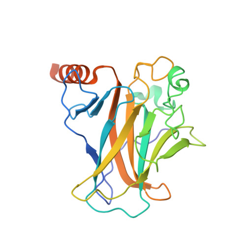
    5A7B

  • PubMed Abstract: 

    Bioisosteric replacements are widely used in medicinal chemistry to improve physicochemical and ADME properties of molecules while retaining or improving affinity. Here, using the p53 cancer mutant Y220C as a test case, we investigate both computationally and experimentally whether an ethynyl moiety is a suitable bioisostere to replace iodine in ligands that form halogen bonds with the protein backbone. This bioisosteric transformation is synthetically feasible via Sonogashira cross-coupling. In our test case of a particularly strong halogen bond, replacement of the iodine with an ethynyl group resulted in a 13-fold affinity loss. High-resolution crystal structures of the two analogues in complex with the p53-Y220C mutant enabled us to correlate the different affinities with particular features of the binding site and subtle changes in ligand binding mode. In addition, using QM calculations and analyzing the PDB, we provide general guidelines for identifying cases where such a transformation is likely to improve ligand recognition.


  • Organizational Affiliation

    MRC Laboratory of Molecular Biology , Francis Crick Avenue, Cambridge CB2 0QH, United Kingdom.


Macromolecules
Find similar proteins by:  (by identity cutoff)  |  3D Structure
Entity ID: 1
MoleculeChains Sequence LengthOrganismDetailsImage
CELLULAR TUMOR ANTIGEN P53
A, B
219Homo sapiensMutation(s): 5 
UniProt & NIH Common Fund Data Resources
Find proteins for P04637 (Homo sapiens)
Explore P04637 
Go to UniProtKB:  P04637
PHAROS:  P04637
GTEx:  ENSG00000141510 
Entity Groups  
Sequence Clusters30% Identity50% Identity70% Identity90% Identity95% Identity100% Identity
UniProt GroupP04637
Sequence Annotations
Expand
  • Reference Sequence
Small Molecules
Binding Affinity Annotations 
IDSourceBinding Affinity
KMN Binding MOAD:  5A7B Kd: 2.71e+5 (nM) from 1 assay(s)
Experimental Data & Validation

Experimental Data

  • Method: X-RAY DIFFRACTION
  • Resolution: 1.40 Å
  • R-Value Free: 0.192 
  • R-Value Work: 0.176 
  • R-Value Observed: 0.177 
  • Space Group: P 21 21 21
Unit Cell:
Length ( Å )Angle ( ˚ )
a = 65.12α = 90
b = 71.111β = 90
c = 105.114γ = 90
Software Package:
Software NamePurpose
PHENIXrefinement
PHENIXphasing

Structure Validation

View Full Validation Report


Ligand Structure Quality Assessment 


Entry History 

Deposition Data

Revision History  (Full details and data files)

  • Version 1.0: 2015-09-30
    Type: Initial release
  • Version 1.1: 2016-01-13
    Changes: Database references
  • Version 1.2: 2019-05-08
    Changes: Data collection, Experimental preparation, Other
  • Version 1.3: 2019-05-15
    Changes: Data collection, Experimental preparation
  • Version 1.4: 2024-01-10
    Changes: Data collection, Database references, Derived calculations, Other, Refinement description