4YLS

Tubulin Glutamylase


Experimental Data Snapshot

  • Method: X-RAY DIFFRACTION
  • Resolution: 2.60 Å
  • R-Value Free: 0.246 
  • R-Value Work: 0.210 
  • R-Value Observed: 0.212 

wwPDB Validation   3D Report Full Report


Ligand Structure Quality Assessment 


This is version 1.3 of the entry. See complete history


Literature

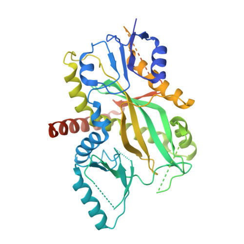
Multivalent Microtubule Recognition by Tubulin Tyrosine Ligase-like Family Glutamylases.

Garnham, C.P.Vemu, A.Wilson-Kubalek, E.M.Yu, I.Szyk, A.Lander, G.C.Milligan, R.A.Roll-Mecak, A.

(2015) Cell 161: 1112-1123

  • DOI: https://doi.org/10.1016/j.cell.2015.04.003
  • Primary Citation of Related Structures:  
    4YLR, 4YLS

  • PubMed Abstract: 

    Glutamylation, the most prevalent tubulin posttranslational modification, marks stable microtubules and regulates recruitment and activity of microtubule- interacting proteins. Nine enzymes of the tubulin tyrosine ligase-like (TTLL) family catalyze glutamylation. TTLL7, the most abundant neuronal glutamylase, adds glutamates preferentially to the β-tubulin tail. Coupled with ensemble and single-molecule biochemistry, our hybrid X-ray and cryo-electron microscopy structure of TTLL7 bound to the microtubule delineates a tripartite microtubule recognition strategy. The enzyme uses its core to engage the disordered anionic tails of α- and β-tubulin, and a flexible cationic domain to bind the microtubule and position itself for β-tail modification. Furthermore, we demonstrate that all single-chain TTLLs with known glutamylase activity utilize a cationic microtubule-binding domain analogous to that of TTLL7. Therefore, our work reveals the combined use of folded and intrinsically disordered substrate recognition elements as the molecular basis for specificity among the enzymes primarily responsible for chemically diversifying cellular microtubules.


  • Organizational Affiliation

    Cell Biology and Biophysics Unit, Porter Neuroscience Research Center, National Institute of Neurological Disorders and Stroke, Bethesda, MD 20892, USA.


Macromolecules
Find similar proteins by:  (by identity cutoff)  |  3D Structure
Entity ID: 1
MoleculeChains Sequence LengthOrganismDetailsImage
Tubulin polyglutamylase TTLL7487Homo sapiensMutation(s): 1 
Gene Names: TTLL7
EC: 6
UniProt & NIH Common Fund Data Resources
Find proteins for Q6ZT98 (Homo sapiens)
Explore Q6ZT98 
Go to UniProtKB:  Q6ZT98
PHAROS:  Q6ZT98
GTEx:  ENSG00000137941 
Entity Groups  
Sequence Clusters30% Identity50% Identity70% Identity90% Identity95% Identity100% Identity
UniProt GroupQ6ZT98
Sequence Annotations
Expand
  • Reference Sequence
Small Molecules
Ligands 1 Unique
IDChains Name / Formula / InChI Key2D Diagram3D Interactions
ANP
Query on ANP

Download Ideal Coordinates CCD File 
B [auth A]PHOSPHOAMINOPHOSPHONIC ACID-ADENYLATE ESTER
C10 H17 N6 O12 P3
PVKSNHVPLWYQGJ-KQYNXXCUSA-N
Experimental Data & Validation

Experimental Data

  • Method: X-RAY DIFFRACTION
  • Resolution: 2.60 Å
  • R-Value Free: 0.246 
  • R-Value Work: 0.210 
  • R-Value Observed: 0.212 
  • Space Group: C 2 2 21
Unit Cell:
Length ( Å )Angle ( ˚ )
a = 79.126α = 90
b = 122.324β = 90
c = 129.445γ = 90
Software Package:
Software NamePurpose
PHENIXrefinement
HKL-2000data reduction
SCALEPACKdata scaling
PHENIXphasing
PDB_EXTRACTdata extraction

Structure Validation

View Full Validation Report


Ligand Structure Quality Assessment 


Entry History & Funding Information

Deposition Data


Funding OrganizationLocationGrant Number
National Institutes of Health/National Institute of General Medical Sciences (NIH/NIGMS)United States--

Revision History  (Full details and data files)

  • Version 1.0: 2015-06-17
    Type: Initial release
  • Version 1.1: 2017-09-20
    Changes: Author supporting evidence, Derived calculations, Source and taxonomy
  • Version 1.2: 2019-12-25
    Changes: Author supporting evidence
  • Version 1.3: 2024-02-28
    Changes: Data collection, Database references