4X34

Crystal structure of the 53BP1 tandem tudor domain in complex with p53K381acK382me2


Experimental Data Snapshot

  • Method: X-RAY DIFFRACTION
  • Resolution: 1.80 Å
  • R-Value Free: 0.218 
  • R-Value Work: 0.187 
  • R-Value Observed: 0.188 

wwPDB Validation   3D Report Full Report


This is version 1.5 of the entry. See complete history


Literature

An Acetyl-Methyl Switch Drives a Conformational Change in p53.

Tong, Q.Mazur, S.J.Rincon-Arano, H.Rothbart, S.B.Kuznetsov, D.M.Cui, G.Liu, W.H.Gete, Y.Klein, B.J.Jenkins, L.Mer, G.Kutateladze, A.G.Strahl, B.D.Groudine, M.Appella, E.Kutateladze, T.G.

(2015) Structure 23: 322-331

  • DOI: https://doi.org/10.1016/j.str.2014.12.010
  • Primary Citation of Related Structures:  
    4X34

  • PubMed Abstract: 

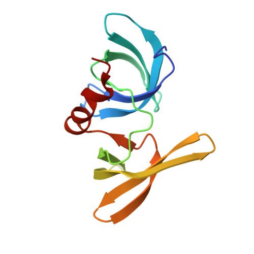
    Individual posttranslational modifications (PTMs) of p53 mediate diverse p53-dependent responses; however, much less is known about the combinatorial action of adjacent modifications. Here, we describe crosstalk between the early DNA damage response mark p53K382me2 and the surrounding PTMs that modulate binding of p53 cofactors, including 53BP1 and p300. The 1.8 Å resolution crystal structure of the tandem Tudor domain (TTD) of 53BP1 in complex with p53 peptide acetylated at K381 and dimethylated at K382 (p53K381acK382me2) reveals that the dual PTM induces a conformational change in p53. The α-helical fold of p53K381acK382me2 positions the side chains of R379, K381ac, and K382me2 to interact with TTD concurrently, reinforcing a modular design of double PTM mimetics. Biochemical and nuclear magnetic resonance analyses show that other surrounding PTMs, including phosphorylation of serine/threonine residues of p53, affect association with TTD. Our findings suggest a novel PTM-driven conformation switch-like mechanism that may regulate p53 interactions with binding partners.


  • Organizational Affiliation

    Department of Pharmacology, University of Colorado School of Medicine, Aurora, CO 80045, USA.


Macromolecules
Find similar proteins by:  (by identity cutoff)  |  3D Structure
Entity ID: 1
MoleculeChains Sequence LengthOrganismDetailsImage
Tumor suppressor p53-binding protein 1
A, B
120Homo sapiensMutation(s): 0 
Gene Names: TP53BP1
UniProt & NIH Common Fund Data Resources
Find proteins for Q12888 (Homo sapiens)
Explore Q12888 
Go to UniProtKB:  Q12888
PHAROS:  Q12888
GTEx:  ENSG00000067369 
Entity Groups  
Sequence Clusters30% Identity50% Identity70% Identity90% Identity95% Identity100% Identity
UniProt GroupQ12888
Sequence Annotations
Expand
  • Reference Sequence

Find similar proteins by:  Sequence   |   3D Structure  

Entity ID: 2
MoleculeChains Sequence LengthOrganismDetailsImage
THR-SER-ARG-HIS-ALY-MLY-LEU-MET-PHE-LYS
C, D
10Homo sapiensMutation(s): 0 
UniProt & NIH Common Fund Data Resources
Find proteins for P04637 (Homo sapiens)
Explore P04637 
Go to UniProtKB:  P04637
PHAROS:  P04637
GTEx:  ENSG00000141510 
Entity Groups  
Sequence Clusters30% Identity50% Identity70% Identity90% Identity95% Identity100% Identity
UniProt GroupP04637
Sequence Annotations
Expand
  • Reference Sequence
Small Molecules
Modified Residues  2 Unique
IDChains TypeFormula2D DiagramParent
ALY
Query on ALY
C, D
L-PEPTIDE LINKINGC8 H16 N2 O3LYS
MLY
Query on MLY
C, D
L-PEPTIDE LINKINGC8 H18 N2 O2LYS
Experimental Data & Validation

Experimental Data

  • Method: X-RAY DIFFRACTION
  • Resolution: 1.80 Å
  • R-Value Free: 0.218 
  • R-Value Work: 0.187 
  • R-Value Observed: 0.188 
  • Space Group: C 2 2 21
Unit Cell:
Length ( Å )Angle ( ˚ )
a = 58.891α = 90
b = 110.939β = 90
c = 96.915γ = 90
Software Package:
Software NamePurpose
HKL-2000data reduction
MOLREPphasing
PHENIXrefinement
PDB_EXTRACTdata extraction
HKL-2000data scaling
MOLREPphasing

Structure Validation

View Full Validation Report


Entry History & Funding Information

Deposition Data


Funding OrganizationLocationGrant Number
National Institutes of Health/National Institute of General Medical Sciences (NIH/NIGMS)United StatesGM101664 (T.G.K.)

Revision History  (Full details and data files)

  • Version 1.0: 2015-03-04
    Type: Initial release
  • Version 1.1: 2015-09-09
    Changes: Data collection
  • Version 1.2: 2017-09-06
    Changes: Author supporting evidence, Derived calculations
  • Version 1.3: 2019-12-25
    Changes: Author supporting evidence, Database references
  • Version 1.4: 2023-09-27
    Changes: Data collection, Database references, Refinement description
  • Version 1.5: 2023-11-15
    Changes: Data collection