4V2O

Structure of saposin B in complex with chloroquine


Experimental Data Snapshot

  • Method: X-RAY DIFFRACTION
  • Resolution: 2.13 Å
  • R-Value Free: 0.254 
  • R-Value Work: 0.229 
  • R-Value Observed: 0.230 

wwPDB Validation   3D Report Full Report


Ligand Structure Quality Assessment 


This is version 1.2 of the entry. See complete history


Literature

The Lysosomal Protein Saposin B Binds Chloroquine.

Huta, B.P.Mehlenbacher, M.R.Nie, Y.Lai, X.Zubieta, C.Bou-Abdallah, F.Doyle, R.P.

(2016) ChemMedChem 11: 277

  • DOI: https://doi.org/10.1002/cmdc.201500494
  • Primary Citation of Related Structures:  
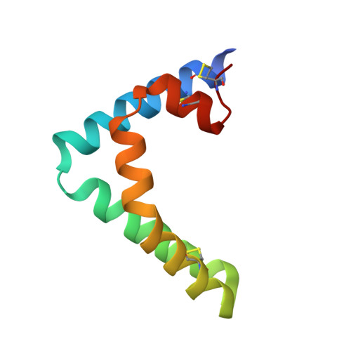
    4V2O

  • PubMed Abstract: 

    Chloroquine (CQ) has been widely used in the treatment of malaria since the 1950s, though toxicity and resistance is increasingly limiting its use in the clinic. More recently, CQ is also becoming recognized as an important therapeutic compound for the treatment of autoimmune disorders and has shown activity as an anticancer agent. However, the full extent of CQ pharmacology in humans is still unclear. Herein, we demonstrate that the lysosomal protein saposin B (sapB), critical for select lipid degradation, binds CQ with implications for both CQ function and toxicity. Using isothermal titration calorimetry (ITC) and fluorescence quenching experiments, CQ was shown to bind to the dimeric form of sapB at both pH 5.5 and pH 7.4 with an average binding affinity of 2.3×10(4)  m(-1). X-ray crystallography confirmed this, and the first complete crystal structure of sapB with a bound small molecule (CQ) is reported. The results suggest that sapB might play a role in mitigating CQ-based toxicity and that sapB might itself be overwhelmed by CQ causing impaired lipid degradation.


  • Organizational Affiliation

    Department of Chemistry, Syracuse University, 111 College Place, Syracuse, NY, 13244, USA.


Macromolecules
Find similar proteins by:  (by identity cutoff)  |  3D Structure
Entity ID: 1
MoleculeChains Sequence LengthOrganismDetailsImage
SAPOSIN-B
A, B, C
82Homo sapiensMutation(s): 0 
Membrane Entity: Yes 
UniProt & NIH Common Fund Data Resources
Find proteins for P07602 (Homo sapiens)
Explore P07602 
Go to UniProtKB:  P07602
PHAROS:  P07602
GTEx:  ENSG00000197746 
Entity Groups  
Sequence Clusters30% Identity50% Identity70% Identity90% Identity95% Identity100% Identity
UniProt GroupP07602
Sequence Annotations
Expand
  • Reference Sequence
Small Molecules
Ligands 1 Unique
IDChains Name / Formula / InChI Key2D Diagram3D Interactions
CLQ
Query on CLQ

Download Ideal Coordinates CCD File 
D [auth B],
E [auth C]
N4-(7-CHLORO-QUINOLIN-4-YL)-N1,N1-DIETHYL-PENTANE-1,4-DIAMINE
C18 H26 Cl N3
WHTVZRBIWZFKQO-CQSZACIVSA-N
Binding Affinity Annotations 
IDSourceBinding Affinity
CLQ Binding MOAD:  4V2O Ka: 3.14e+4 (M^-1) from 1 assay(s)
Experimental Data & Validation

Experimental Data

  • Method: X-RAY DIFFRACTION
  • Resolution: 2.13 Å
  • R-Value Free: 0.254 
  • R-Value Work: 0.229 
  • R-Value Observed: 0.230 
  • Space Group: P 32 2 1
Unit Cell:
Length ( Å )Angle ( ˚ )
a = 75.299α = 90
b = 75.299β = 90
c = 94.716γ = 120
Software Package:
Software NamePurpose
BUSTERrefinement
XDSdata reduction
XSCALEdata scaling

Structure Validation

View Full Validation Report


Ligand Structure Quality Assessment 


Entry History 

Deposition Data

Revision History  (Full details and data files)

  • Version 1.0: 2015-12-09
    Type: Initial release
  • Version 1.1: 2016-03-02
    Changes: Database references
  • Version 1.2: 2024-01-10
    Changes: Data collection, Database references, Derived calculations, Other, Refinement description