4UIP

The complex structure of extracellular domain of EGFR with Repebody (rAC1).


Experimental Data Snapshot

  • Method: X-RAY DIFFRACTION
  • Resolution: 2.95 Å
  • R-Value Free: 0.297 
  • R-Value Work: 0.237 
  • R-Value Observed: 0.240 

wwPDB Validation   3D Report Full Report


Ligand Structure Quality Assessment 


This is version 2.1 of the entry. See complete history


Literature

Enzymatic Prenylation and Oxime Ligation for the Synthesis of Stable and Homogeneous Protein-Drug Conjugates for Targeted Therapy.

Lee, J.Choi, H.Yun, M.Kang, Y.Jung, J.Ryu, Y.Kim, T.Y.Cha, Y.Cho, H.Min, J.Chung, C.Kim, H.

(2015) Angew Chem Int Ed Engl 54: 12020

  • DOI: https://doi.org/10.1002/anie.201505964
  • Primary Citation of Related Structures:  
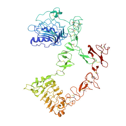
    4UIP

  • PubMed Abstract: 

    Targeted therapy based on protein-drug conjugates has attracted significant attention owing to its high efficacy and low side effects. However, efficient and stable drug conjugation to a protein binder remains a challenge. Herein, a chemoenzymatic method to generate highly stable and homogenous drug conjugates with high efficiency is presented. The approach comprises the insertion of the CaaX sequence at the C-terminal end of the protein binder, prenylation using farnesyltransferase, and drug conjugation through an oxime ligation reaction. MMAF and an EGFR-specific repebody are used as the antitumor agent and protein binder, respectively. The method enables the precisely controlled synthesis of repebody-drug conjugates with high yield and homogeneity. The utility of this approach is illustrated by the notable stability of the repebody-drug conjugates in human plasma, negligible off-target effects, and a remarkable antitumor activity in vivo. The present method can be widely used for generating highly homogeneous and stable PDCs for targeted therapy.


  • Organizational Affiliation

    Department of Biological Sciences, Korea Advanced Institute of Science and Technology (KAIST), Daejeon (Korea).


Macromolecules
Find similar proteins by:  (by identity cutoff)  |  3D Structure
Entity ID: 1
MoleculeChains Sequence LengthOrganismDetailsImage
EPIDERMAL GROWTH FACTOR RECEPTOR618Homo sapiensMutation(s): 0 
UniProt & NIH Common Fund Data Resources
Find proteins for P00533 (Homo sapiens)
Explore P00533 
Go to UniProtKB:  P00533
PHAROS:  P00533
GTEx:  ENSG00000146648 
Entity Groups  
Sequence Clusters30% Identity50% Identity70% Identity90% Identity95% Identity100% Identity
UniProt GroupP00533
Sequence Annotations
Expand
  • Reference Sequence
Find similar proteins by:  (by identity cutoff)  |  3D Structure
Entity ID: 2
MoleculeChains Sequence LengthOrganismDetailsImage
REPEBODY (RAC1)251Listeria monocytogenessynthetic constructEptatretus burgeri
This entity is chimeric
Mutation(s): 0 
UniProt
Find proteins for Q4G1L3 (Eptatretus burgeri)
Explore Q4G1L3 
Go to UniProtKB:  Q4G1L3
Find proteins for E0ACT6 (Listeria monocytogenes)
Explore E0ACT6 
Go to UniProtKB:  E0ACT6
Entity Groups  
Sequence Clusters30% Identity50% Identity70% Identity90% Identity95% Identity100% Identity
UniProt GroupsE0ACT6Q4G1L3
Sequence Annotations
Expand
  • Reference Sequence
Oligosaccharides

Help

Entity ID: 3
MoleculeChains Length2D Diagram Glycosylation3D Interactions
2-acetamido-2-deoxy-beta-D-glucopyranose-(1-4)-2-acetamido-2-deoxy-beta-D-glucopyranose
C
2N-Glycosylation
Glycosylation Resources
GlyTouCan:  G42666HT
GlyCosmos:  G42666HT
GlyGen:  G42666HT
Small Molecules
Ligands 1 Unique
IDChains Name / Formula / InChI Key2D Diagram3D Interactions
NAG
Query on NAG

Download Ideal Coordinates CCD File 
D [auth A],
E [auth A]
2-acetamido-2-deoxy-beta-D-glucopyranose
C8 H15 N O6
OVRNDRQMDRJTHS-FMDGEEDCSA-N
Experimental Data & Validation

Experimental Data

  • Method: X-RAY DIFFRACTION
  • Resolution: 2.95 Å
  • R-Value Free: 0.297 
  • R-Value Work: 0.237 
  • R-Value Observed: 0.240 
  • Space Group: P 2 21 21
Unit Cell:
Length ( Å )Angle ( ˚ )
a = 66.79α = 90
b = 88.34β = 90
c = 189.21γ = 90
Software Package:
Software NamePurpose
REFMACrefinement

Structure Validation

View Full Validation Report


Ligand Structure Quality Assessment 


Entry History 

Deposition Data

Revision History  (Full details and data files)

  • Version 1.0: 2015-11-25
    Type: Initial release
  • Version 1.1: 2015-12-23
    Changes: Structure summary
  • Version 1.2: 2017-03-15
    Changes: Source and taxonomy
  • Version 2.0: 2020-07-29
    Type: Remediation
    Reason: Carbohydrate remediation
    Changes: Advisory, Atomic model, Data collection, Derived calculations, Other, Structure summary
  • Version 2.1: 2024-01-10
    Changes: Data collection, Database references, Refinement description, Structure summary