4TKQ

Native-SAD phasing for YetJ from Bacillus Subtilis


Experimental Data Snapshot

  • Method: X-RAY DIFFRACTION
  • Resolution: 2.80 Å
  • R-Value Free: 0.260 
  • R-Value Work: 0.221 
  • R-Value Observed: 0.223 

wwPDB Validation   3D Report Full Report


This is version 1.5 of the entry. See complete history


Literature

Multi-crystal native SAD analysis at 6 keV.

Liu, Q.Guo, Y.Chang, Y.Cai, Z.Assur, Z.Mancia, F.Greene, M.I.Hendrickson, W.A.

(2014) Acta Crystallogr D Biol Crystallogr 70: 2544-2557

  • DOI: https://doi.org/10.1107/S1399004714013376
  • Primary Citation of Related Structures:  
    4TKQ, 4TKR, 4TKS

  • PubMed Abstract: 

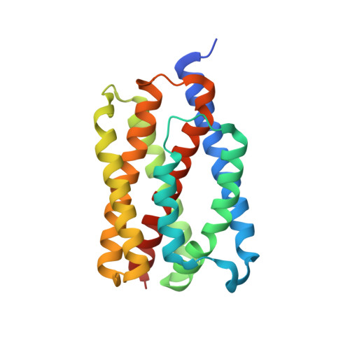
    Anomalous diffraction signals from typical native macromolecules are very weak, frustrating their use in de novo structure determination. Here, native SAD procedures are described to enhance signal to noise in anomalous diffraction by using multiple crystals in combination with synchrotron X-rays at 6 keV. Increased anomalous signals were obtained at 6 keV compared with 7 keV X-ray energy, which was used for previous native SAD analyses. A feasibility test of multi-crystal-based native SAD phasing was performed at 3.2 Å resolution for a known tyrosine protein kinase domain, and real-life applications were made to two novel membrane proteins at about 3.0 Å resolution. The three applications collectively serve to validate the robust feasibility of native SAD phasing at lower energy.


  • Organizational Affiliation

    NYCOMPS, New York Structural Biology Center, New York, NY 10032, USA.


Macromolecules
Find similar proteins by:  (by identity cutoff)  |  3D Structure
Entity ID: 1
MoleculeChains Sequence LengthOrganismDetailsImage
Uncharacterized protein YetJ217Bacillus subtilis subsp. subtilis str. 168Mutation(s): 0 
Gene Names: yetJBSU07200
Membrane Entity: Yes 
UniProt
Find proteins for O31539 (Bacillus subtilis (strain 168))
Explore O31539 
Go to UniProtKB:  O31539
Entity Groups  
Sequence Clusters30% Identity50% Identity70% Identity90% Identity95% Identity100% Identity
UniProt GroupO31539
Sequence Annotations
Expand
  • Reference Sequence
Experimental Data & Validation

Experimental Data

  • Method: X-RAY DIFFRACTION
  • Resolution: 2.80 Å
  • R-Value Free: 0.260 
  • R-Value Work: 0.221 
  • R-Value Observed: 0.223 
  • Space Group: P 65 2 2
Unit Cell:
Length ( Å )Angle ( ˚ )
a = 61.869α = 90
b = 61.869β = 90
c = 288.401γ = 120
Software Package:
Software NamePurpose
PHENIXrefinement

Structure Validation

View Full Validation Report


Entry History & Funding Information

Deposition Data


Funding OrganizationLocationGrant Number
National Institutes of Health/National Institute of General Medical Sciences (NIH/NIGMS)United StatesGM095315

Revision History  (Full details and data files)

  • Version 1.0: 2014-06-18
    Type: Initial release
  • Version 1.1: 2014-10-08
    Changes: Database references
  • Version 1.2: 2014-10-22
    Changes: Database references
  • Version 1.3: 2017-09-06
    Changes: Author supporting evidence, Derived calculations, Other, Source and taxonomy
  • Version 1.4: 2019-12-25
    Changes: Author supporting evidence
  • Version 1.5: 2023-12-27
    Changes: Data collection, Database references, Refinement description