4RIX

Crystal structure of an EGFR/HER3 kinase domain heterodimer containing the cancer-associated HER3-Q790R mutation


Experimental Data Snapshot

  • Method: X-RAY DIFFRACTION
  • Resolution: 3.10 Å
  • R-Value Free: 0.258 
  • R-Value Work: 0.207 
  • R-Value Observed: 0.210 

wwPDB Validation   3D Report Full Report


Ligand Structure Quality Assessment 


This is version 1.2 of the entry. See complete history


Literature

Structural analysis of the EGFR/HER3 heterodimer reveals the molecular basis for activating HER3 mutations.

Littlefield, P.Liu, L.Mysore, V.Shan, Y.Shaw, D.E.Jura, N.

(2014) Sci Signal 7: ra114-ra114

  • DOI: https://doi.org/10.1126/scisignal.2005786
  • Primary Citation of Related Structures:  
    4RIW, 4RIX, 4RIY

  • PubMed Abstract: 

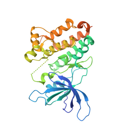
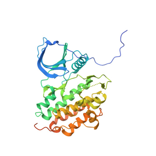
    The human epidermal growth factor receptor (HER) tyrosine kinases homo- and heterodimerize to activate downstream signaling pathways. HER3 is a catalytically impaired member of the HER family that contributes to the development of several human malignancies and is mutated in a subset of cancers. HER3 signaling depends on heterodimerization with a catalytically active partner, in particular epidermal growth factor receptor (EGFR) (the founding family member, also known as HER1) or HER2. The activity of homodimeric complexes of catalytically active HER family members depends on allosteric activation between the two kinase domains. To determine the structural basis for HER3 signaling through heterodimerization with a catalytically active HER family member, we solved the crystal structure of the heterodimeric complex formed by the isolated kinase domains of EGFR and HER3. The structure showed HER3 as an allosteric activator of EGFR and revealed a conserved role of the allosteric mechanism in activation of HER family members through heterodimerization. To understand the effects of cancer-associated HER3 mutations at the molecular level, we solved the structures of two kinase domains of HER3 mutants, each in a heterodimeric complex with the kinase domain of EGFR. These structures, combined with biochemical analysis and molecular dynamics simulations, indicated that the cancer-associated HER3 mutations enhanced the allosteric activator function of HER3 by redesigning local interactions at the dimerization interface.


  • Organizational Affiliation

    Cardiovascular Research Institute, University of California San Francisco, San Francisco, CA 94158, USA.


Macromolecules
Find similar proteins by:  (by identity cutoff)  |  3D Structure
Entity ID: 1
MoleculeChains Sequence LengthOrganismDetailsImage
Receptor tyrosine-protein kinase erbB-3
A, C
326Homo sapiensMutation(s): 1 
Gene Names: ERBB3HER3
EC: 2.7.10.1
UniProt & NIH Common Fund Data Resources
Find proteins for P21860 (Homo sapiens)
Explore P21860 
Go to UniProtKB:  P21860
PHAROS:  P21860
GTEx:  ENSG00000065361 
Entity Groups  
Sequence Clusters30% Identity50% Identity70% Identity90% Identity95% Identity100% Identity
UniProt GroupP21860
Sequence Annotations
Expand
  • Reference Sequence
Find similar proteins by:  (by identity cutoff)  |  3D Structure
Entity ID: 2
MoleculeChains Sequence LengthOrganismDetailsImage
Epidermal growth factor receptor
B, D
345Homo sapiensMutation(s): 3 
Gene Names: EGFR
EC: 2.7.10.1
UniProt & NIH Common Fund Data Resources
Find proteins for P00533 (Homo sapiens)
Explore P00533 
Go to UniProtKB:  P00533
PHAROS:  P00533
GTEx:  ENSG00000146648 
Entity Groups  
Sequence Clusters30% Identity50% Identity70% Identity90% Identity95% Identity100% Identity
UniProt GroupP00533
Sequence Annotations
Expand
  • Reference Sequence
Small Molecules
Ligands 3 Unique
IDChains Name / Formula / InChI Key2D Diagram3D Interactions
ANP
Query on ANP

Download Ideal Coordinates CCD File 
E [auth A],
I [auth C]
PHOSPHOAMINOPHOSPHONIC ACID-ADENYLATE ESTER
C10 H17 N6 O12 P3
PVKSNHVPLWYQGJ-KQYNXXCUSA-N
ADP
Query on ADP

Download Ideal Coordinates CCD File 
G [auth B],
K [auth D]
ADENOSINE-5'-DIPHOSPHATE
C10 H15 N5 O10 P2
XTWYTFMLZFPYCI-KQYNXXCUSA-N
MG
Query on MG

Download Ideal Coordinates CCD File 
F [auth A],
H [auth B],
J [auth C],
L [auth D]
MAGNESIUM ION
Mg
JLVVSXFLKOJNIY-UHFFFAOYSA-N
Experimental Data & Validation

Experimental Data

  • Method: X-RAY DIFFRACTION
  • Resolution: 3.10 Å
  • R-Value Free: 0.258 
  • R-Value Work: 0.207 
  • R-Value Observed: 0.210 
  • Space Group: P 1 21 1
Unit Cell:
Length ( Å )Angle ( ˚ )
a = 64.649α = 90
b = 155.053β = 111.09
c = 86.857γ = 90
Software Package:
Software NamePurpose
Blu-Icedata collection
PHASERphasing
PHENIXrefinement
MOSFLMdata reduction
SCALAdata scaling

Structure Validation

View Full Validation Report


Ligand Structure Quality Assessment 


Entry History 

Deposition Data

Revision History  (Full details and data files)

  • Version 1.0: 2014-12-10
    Type: Initial release
  • Version 1.1: 2014-12-17
    Changes: Database references
  • Version 1.2: 2023-09-20
    Changes: Data collection, Database references, Derived calculations, Refinement description