4RFZ

Crystal structure of BTK kinase domain complexed with 6-(dimethylamino)-8-fluoro-2-[2-(hydroxymethyl)-3-[1-methyl-5-[[5-(morpholine-4-carbonyl)-2-pyridyl]amino]-6-oxo-3-pyridyl]phenyl]isoquinolin-1-one


Experimental Data Snapshot

  • Method: X-RAY DIFFRACTION
  • Resolution: 1.17 Å
  • R-Value Free: 0.184 
  • R-Value Work: 0.157 
  • R-Value Observed: 0.158 

wwPDB Validation   3D Report Full Report


Ligand Structure Quality Assessment 


This is version 1.3 of the entry. See complete history


Literature

Finding the perfect spot for fluorine: Improving potency up to 40-fold during a rational fluorine scan of a Bruton's Tyrosine Kinase (BTK) inhibitor scaffold.

Lou, Y.Sweeney, Z.K.Kuglstatter, A.Davis, D.Goldstein, D.M.Han, X.Hong, J.Kocer, B.Kondru, R.K.Litman, R.McIntosh, J.Sarma, K.Suh, J.Taygerly, J.Owens, T.D.

(2015) Bioorg Med Chem Lett 25: 367-371

  • DOI: https://doi.org/10.1016/j.bmcl.2014.11.030
  • Primary Citation of Related Structures:  
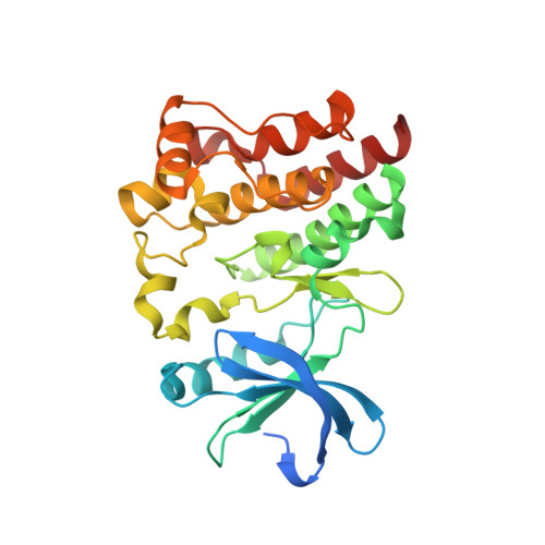
    4RFY, 4RFZ, 4RG0

  • PubMed Abstract: 

    A rational fluorine scan based on co-crystal structures was explored to increase the potency of a series of selective BTK inhibitors. While fluorine substitution on a saturated bicyclic ring system yields no apparent benefit, the same operation on an unsaturated bicyclic ring can increase HWB activity by up to 40-fold. Comparison of co-crystal structures of parent molecules and fluorinated counterparts revealed the importance of placing fluorine at the optimal position to achieve favorable interactions with protein side chains.


  • Organizational Affiliation

    Hoffmann-La Roche Inc., pRED, Pharma Research & Early Development, Small Molecule Research, Discovery Chemistry, 3431 Hillview Ave, Palo Alto, CA 94304, United States. Electronic address: ylou@nurix-inc.com.


Macromolecules
Find similar proteins by:  (by identity cutoff)  |  3D Structure
Entity ID: 1
MoleculeChains Sequence LengthOrganismDetailsImage
Tyrosine-protein kinase BTK283Homo sapiensMutation(s): 4 
Gene Names: BTKAGMX1ATKBPK
EC: 2.7.10.2
UniProt & NIH Common Fund Data Resources
Find proteins for Q06187 (Homo sapiens)
Explore Q06187 
Go to UniProtKB:  Q06187
PHAROS:  Q06187
GTEx:  ENSG00000010671 
Entity Groups  
Sequence Clusters30% Identity50% Identity70% Identity90% Identity95% Identity100% Identity
UniProt GroupQ06187
Sequence Annotations
Expand
  • Reference Sequence
Small Molecules
Ligands 2 Unique
IDChains Name / Formula / InChI Key2D Diagram3D Interactions
3OV
Query on 3OV

Download Ideal Coordinates CCD File 
B [auth A]6-(dimethylamino)-8-fluoro-2-[2-(hydroxymethyl)-3-(1-methyl-5-{[5-(morpholin-4-ylcarbonyl)pyridin-2-yl]amino}-6-oxo-1,6-dihydropyridin-3-yl)phenyl]isoquinolin-1(2H)-one
C34 H33 F N6 O5
PCVMXQPADBQRFE-UHFFFAOYSA-N
DMS
Query on DMS

Download Ideal Coordinates CCD File 
C [auth A],
D [auth A]
DIMETHYL SULFOXIDE
C2 H6 O S
IAZDPXIOMUYVGZ-UHFFFAOYSA-N
Binding Affinity Annotations 
IDSourceBinding Affinity
3OV Binding MOAD:  4RFZ IC50: 40 (nM) from 1 assay(s)
BindingDB:  4RFZ IC50: 40 (nM) from 1 assay(s)
Experimental Data & Validation

Experimental Data

  • Method: X-RAY DIFFRACTION
  • Resolution: 1.17 Å
  • R-Value Free: 0.184 
  • R-Value Work: 0.157 
  • R-Value Observed: 0.158 
  • Space Group: P 21 21 2
Unit Cell:
Length ( Å )Angle ( ˚ )
a = 71.936α = 90
b = 106.197β = 90
c = 38.22γ = 90
Software Package:
Software NamePurpose
REFMACrefinement
CNSrefinement
ADSCdata collection
DENZOdata reduction
SCALEPACKdata scaling
CNSphasing

Structure Validation

View Full Validation Report


Ligand Structure Quality Assessment 


Entry History 

Deposition Data

Revision History  (Full details and data files)

  • Version 1.0: 2014-12-24
    Type: Initial release
  • Version 1.1: 2015-01-14
    Changes: Database references
  • Version 1.2: 2019-07-17
    Changes: Data collection, Refinement description
  • Version 1.3: 2023-09-20
    Changes: Data collection, Database references, Derived calculations, Refinement description