4NGE

Crystal Structure of Human Presequence Protease in Complex with Amyloid-beta (1-40)


Experimental Data Snapshot

  • Method: X-RAY DIFFRACTION
  • Resolution: 2.70 Å
  • R-Value Free: 0.232 
  • R-Value Work: 0.191 
  • R-Value Observed: 0.193 

wwPDB Validation   3D Report Full Report


This is version 1.3 of the entry. See complete history


Literature

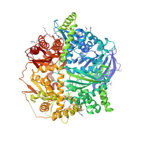
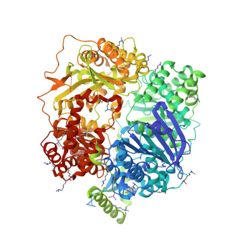
Molecular basis of substrate recognition and degradation by human presequence protease.

King, J.V.Liang, W.G.Scherpelz, K.P.Schilling, A.B.Meredith, S.C.Tang, W.J.

(2014) Structure 22: 996-1007

  • DOI: https://doi.org/10.1016/j.str.2014.05.003
  • Primary Citation of Related Structures:  
    4L3T, 4NGE

  • PubMed Abstract: 

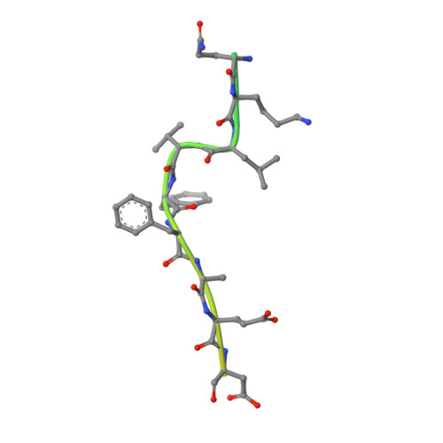
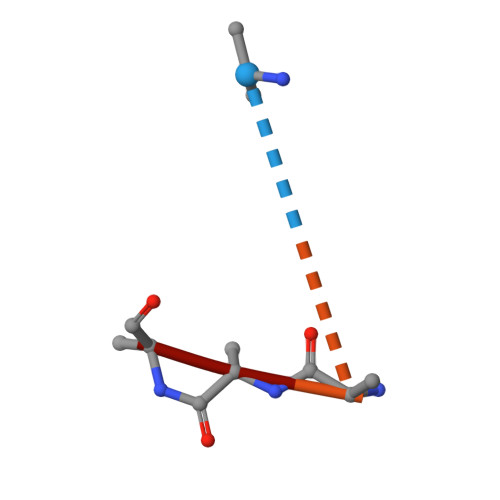
    Human presequence protease (hPreP) is an M16 metalloprotease localized in mitochondria. There, hPreP facilitates proteostasis by utilizing an ∼13,300-Å(3) catalytic chamber to degrade a diverse array of potentially toxic peptides, including mitochondrial presequences and β-amyloid (Aβ), the latter of which contributes to Alzheimer disease pathogenesis. Here, we report crystal structures for hPreP alone and in complex with Aβ, which show that hPreP uses size exclusion and charge complementation for substrate recognition. These structures also reveal hPreP-specific features that permit a diverse array of peptides, with distinct distributions of charged and hydrophobic residues, to be specifically captured, cleaved, and have their amyloidogenic features destroyed. SAXS analysis demonstrates that hPreP in solution exists in dynamic equilibrium between closed and open states, with the former being preferred. Furthermore, Aβ binding induces the closed state and hPreP dimerization. Together, these data reveal the molecular basis for flexible yet specific substrate recognition and degradation by hPreP.


  • Organizational Affiliation

    Ben May Department for Cancer Research, The University of Chicago, 929 E. 57(th) Street, Chicago, IL 60637, USA.


Macromolecules
Find similar proteins by:  (by identity cutoff)  |  3D Structure
Entity ID: 1
MoleculeChains Sequence LengthOrganismDetailsImage
Presequence protease, mitochondrial1,014Homo sapiensMutation(s): 1 
Gene Names: KIAA1104MP1PITRM1
EC: 3.4.24
UniProt & NIH Common Fund Data Resources
Find proteins for Q5JRX3 (Homo sapiens)
Explore Q5JRX3 
Go to UniProtKB:  Q5JRX3
PHAROS:  Q5JRX3
GTEx:  ENSG00000107959 
Entity Groups  
Sequence Clusters30% Identity50% Identity70% Identity90% Identity95% Identity100% Identity
UniProt GroupQ5JRX3
Sequence Annotations
Expand
  • Reference Sequence
Find similar proteins by:  (by identity cutoff)  |  3D Structure
Entity ID: 2
MoleculeChains Sequence LengthOrganismDetailsImage
Beta-amyloid protein 40
B, E
40Homo sapiensMutation(s): 0 
UniProt & NIH Common Fund Data Resources
Find proteins for P05067 (Homo sapiens)
Explore P05067 
Go to UniProtKB:  P05067
PHAROS:  P05067
GTEx:  ENSG00000142192 
Entity Groups  
Sequence Clusters30% Identity50% Identity70% Identity90% Identity95% Identity100% Identity
UniProt GroupP05067
Sequence Annotations
Expand
  • Reference Sequence

Find similar proteins by:  Sequence   |   3D Structure  

Entity ID: 3
MoleculeChains Sequence LengthOrganismDetailsImage
Beta-amyloid protein 40
C, F
7Homo sapiensMutation(s): 0 
Gene Names: KIAA1104MP1PITRM1
EC: 3.4.24
Sequence Annotations
Expand
  • Reference Sequence
Find similar proteins by:  (by identity cutoff)  |  3D Structure
Entity ID: 4
MoleculeChains Sequence LengthOrganismDetailsImage
Presequence protease, mitochondrial1,014Homo sapiensMutation(s): 1 
UniProt & NIH Common Fund Data Resources
Find proteins for Q5JRX3 (Homo sapiens)
Explore Q5JRX3 
Go to UniProtKB:  Q5JRX3
PHAROS:  Q5JRX3
GTEx:  ENSG00000107959 
Entity Groups  
Sequence Clusters30% Identity50% Identity70% Identity90% Identity95% Identity100% Identity
UniProt GroupQ5JRX3
Sequence Annotations
Expand
  • Reference Sequence
Small Molecules
Modified Residues  3 Unique
IDChains TypeFormula2D DiagramParent
CAS
Query on CAS
A
L-PEPTIDE LINKINGC5 H12 As N O2 SCYS
MLY
Query on MLY
A
L-PEPTIDE LINKINGC8 H18 N2 O2LYS
MLZ
Query on MLZ
A
L-PEPTIDE LINKINGC7 H16 N2 O2LYS
Experimental Data & Validation

Experimental Data

  • Method: X-RAY DIFFRACTION
  • Resolution: 2.70 Å
  • R-Value Free: 0.232 
  • R-Value Work: 0.191 
  • R-Value Observed: 0.193 
  • Space Group: C 1 2 1
Unit Cell:
Length ( Å )Angle ( ˚ )
a = 247.275α = 90
b = 86.183β = 127.56
c = 158.611γ = 90
Software Package:
Software NamePurpose
HKL-3000data collection
PHASERphasing
PHENIXrefinement
HKL-3000data reduction
HKL-3000data scaling

Structure Validation

View Full Validation Report


Entry History 

Deposition Data

Revision History  (Full details and data files)

  • Version 1.0: 2014-05-14
    Type: Initial release
  • Version 1.1: 2014-05-21
    Changes: Database references
  • Version 1.2: 2014-07-09
    Changes: Database references
  • Version 1.3: 2014-07-23
    Changes: Database references