4LOV

Crystal structure of FimH in complex with Heptylmannoside


Experimental Data Snapshot

  • Method: X-RAY DIFFRACTION
  • Resolution: 1.50 Å
  • R-Value Free: 0.190 
  • R-Value Work: 0.161 
  • R-Value Observed: 0.163 

wwPDB Validation   3D Report Full Report


Ligand Structure Quality Assessment 


This is version 1.2 of the entry. See complete history


Literature

Study of the Structural and Dynamic Effects in the FimH Adhesin upon alpha-d-Heptyl Mannose Binding.

Vanwetswinkel, S.Volkov, A.N.Sterckx, Y.G.Garcia-Pino, A.Buts, L.Vranken, W.F.Bouckaert, J.Roy, R.Wyns, L.van Nuland, N.A.

(2014) J Med Chem 57: 1416-1427

  • DOI: https://doi.org/10.1021/jm401666c
  • Primary Citation of Related Structures:  
    3ZPD, 4LOV

  • PubMed Abstract: 

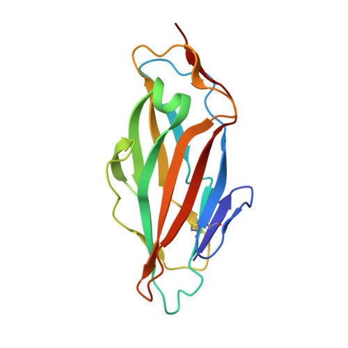
    Uropathogenic Escherichia coli cause urinary tract infections by adhering to mannosylated receptors on the human urothelium via the carbohydrate-binding domain of the FimH adhesin (FimHL). Numerous α-d-mannopyranosides, including α-d-heptyl mannose (HM), inhibit this process by interacting with FimHL. To establish the molecular basis of the high-affinity HM binding, we solved the solution structure of the apo form and the crystal structure of the FimHL-HM complex. NMR relaxation analysis revealed that protein dynamics were not affected by the sugar binding, yet HM addition promoted protein dimerization, which was further confirmed by small-angle X-ray scattering. Finally, to address the role of Y48, part of the "tyrosine gate" believed to govern the affinity and specificity of mannoside binding, we characterized the FimHL Y48A mutant, whose conformational, dynamical, and HM binding properties were found to be very similar to those of the wild-type protein.


  • Organizational Affiliation

    Jean Jeener NMR Centre, Structural Biology Brussels, Vrije Universiteit Brussel , Pleinlaan 2, 1050 Brussels, Belgium.


Macromolecules
Find similar proteins by:  (by identity cutoff)  |  3D Structure
Entity ID: 1
MoleculeChains Sequence LengthOrganismDetailsImage
Protein FimH158Escherichia coliMutation(s): 0 
Gene Names: fimHb4320JW4283
UniProt
Find proteins for P08191 (Escherichia coli (strain K12))
Explore P08191 
Go to UniProtKB:  P08191
Entity Groups  
Sequence Clusters30% Identity50% Identity70% Identity90% Identity95% Identity100% Identity
UniProt GroupP08191
Sequence Annotations
Expand
  • Reference Sequence
Experimental Data & Validation

Experimental Data

  • Method: X-RAY DIFFRACTION
  • Resolution: 1.50 Å
  • R-Value Free: 0.190 
  • R-Value Work: 0.161 
  • R-Value Observed: 0.163 
  • Space Group: P 21 21 21
Unit Cell:
Length ( Å )Angle ( ˚ )
a = 39.9α = 90
b = 41.82β = 90
c = 97.49γ = 90
Software Package:
Software NamePurpose
PHASERphasing
PHENIXrefinement
XDSdata reduction
XDSdata scaling
BUSTERrefinement

Structure Validation

View Full Validation Report


Ligand Structure Quality Assessment 


Entry History 

Deposition Data

Revision History  (Full details and data files)

  • Version 1.0: 2014-02-12
    Type: Initial release
  • Version 1.1: 2014-03-12
    Changes: Database references
  • Version 1.2: 2020-07-29
    Type: Remediation
    Reason: Carbohydrate remediation
    Changes: Derived calculations, Structure summary