4KZO

Crystal Structure Analysis of human IDH1 mutants in complex with NADP+ and Ca2+/alpha-Ketoglutarate


Experimental Data Snapshot

  • Method: X-RAY DIFFRACTION
  • Resolution: 2.20 Å
  • R-Value Free: 0.221 
  • R-Value Work: 0.178 
  • R-Value Observed: 0.180 

wwPDB Validation   3D Report Full Report


Ligand Structure Quality Assessment 


This is version 1.1 of the entry. See complete history


Literature

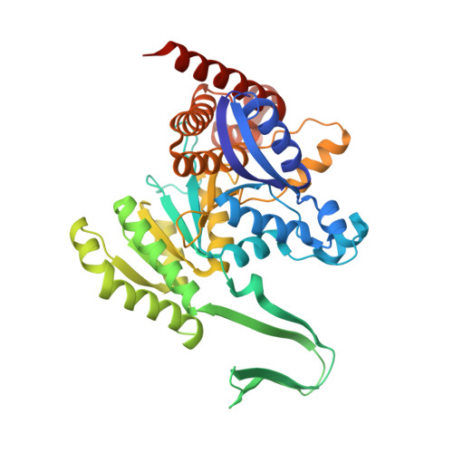
Mutant IDH1 Enhances the Production of 2-Hydroxyglutarate Due to Its Kinetic Mechanism.

Rendina, A.R.Pietrak, B.Smallwood, A.Zhao, H.Qi, H.Quinn, C.Adams, N.D.Concha, N.Duraiswami, C.Thrall, S.H.Sweitzer, S.Schwartz, B.

(2013) Biochemistry 52: 4563-4577

  • DOI: https://doi.org/10.1021/bi400514k
  • Primary Citation of Related Structures:  
    4KZO, 4L03, 4L04, 4L06

  • PubMed Abstract: 

    The human, cytosolic enzyme isocitrate dehydrogenase 1 (IDH1) reversibly converts isocitrate to α-ketoglutarate (αKG). Cancer-associated somatic mutations in IDH1 result in a loss of this normal function but a gain in a new or neomorphic ability to convert αKG to the oncometabolite 2-hydroxyglutarate (2HG). To improve our understanding of the basis for this phenomenon, we have conducted a detailed kinetic study of wild-type IDH1 as well as the known 2HG-producing clinical R132H and G97D mutants and mechanistic Y139D and (newly described) G97N mutants. In the reductive direction of the normal reaction (αKG to isocitrate), dead-end inhibition studies suggest that wild-type IDH1 goes through a random sequential mechanism, similar to previous reports on related mammalian IDH enzymes. However, analogous experiments studying the reductive neomorphic reaction (αKG to 2HG) with the mutant forms of IDH1 are more consistent with an ordered sequential mechanism, with NADPH binding before αKG. This result was further confirmed by primary kinetic isotope effects for which saturating with αKG greatly reduced the observed isotope effect on (D)(V/K)NADPH. For the mutant IDH1 enzyme, the change in mechanism was consistently associated with reduced efficiencies in the use of αKG as a substrate and enhanced efficiencies using NADPH as a substrate. We propose that the sum of these kinetic changes allows the mutant IDH1 enzymes to reductively trap αKG directly into 2HG, rather than allowing it to react with carbon dioxide and form isocitrate, as occurs in the wild-type enzyme.


  • Organizational Affiliation

    Departments of Biological Reagents and Assay Development, Cancer Epigenetics, and Computational and Structural Chemistry, GlaxoSmithKline, 1250 South Collegeville Road, Collegeville, Pennsylvania 19426, USA.


Macromolecules
Find similar proteins by:  (by identity cutoff)  |  3D Structure
Entity ID: 1
MoleculeChains Sequence LengthOrganismDetailsImage
Isocitrate dehydrogenase [NADP] cytoplasmic
A, B, C
425Homo sapiensMutation(s): 1 
Gene Names: IDH1PICD
EC: 1.1.1.42
UniProt & NIH Common Fund Data Resources
Find proteins for O75874 (Homo sapiens)
Explore O75874 
Go to UniProtKB:  O75874
PHAROS:  O75874
GTEx:  ENSG00000138413 
Entity Groups  
Sequence Clusters30% Identity50% Identity70% Identity90% Identity95% Identity100% Identity
UniProt GroupO75874
Sequence Annotations
Expand
  • Reference Sequence
Small Molecules
Ligands 4 Unique
IDChains Name / Formula / InChI Key2D Diagram3D Interactions
NAP
Query on NAP

Download Ideal Coordinates CCD File 
E [auth A],
I [auth B],
L [auth C]
NADP NICOTINAMIDE-ADENINE-DINUCLEOTIDE PHOSPHATE
C21 H28 N7 O17 P3
XJLXINKUBYWONI-NNYOXOHSSA-N
AKG
Query on AKG

Download Ideal Coordinates CCD File 
F [auth A],
J [auth B],
M [auth C]
2-OXOGLUTARIC ACID
C5 H6 O5
KPGXRSRHYNQIFN-UHFFFAOYSA-N
EDO
Query on EDO

Download Ideal Coordinates CCD File 
G [auth A]1,2-ETHANEDIOL
C2 H6 O2
LYCAIKOWRPUZTN-UHFFFAOYSA-N
CA
Query on CA

Download Ideal Coordinates CCD File 
D [auth A],
H [auth B],
K [auth C]
CALCIUM ION
Ca
BHPQYMZQTOCNFJ-UHFFFAOYSA-N
Binding Affinity Annotations 
IDSourceBinding Affinity
NAP Binding MOAD:  4KZO Kd: 43 (nM) from 1 assay(s)
Experimental Data & Validation

Experimental Data

  • Method: X-RAY DIFFRACTION
  • Resolution: 2.20 Å
  • R-Value Free: 0.221 
  • R-Value Work: 0.178 
  • R-Value Observed: 0.180 
  • Space Group: C 2 2 21
Unit Cell:
Length ( Å )Angle ( ˚ )
a = 96.36α = 90
b = 274.598β = 90
c = 115.99γ = 90
Software Package:
Software NamePurpose
PHENIXrefinement
PDB_EXTRACTdata extraction
HKL-2000data reduction
HKL-2000data scaling
PHASERphasing

Structure Validation

View Full Validation Report


Ligand Structure Quality Assessment 


Entry History 

Deposition Data

Revision History  (Full details and data files)

  • Version 1.0: 2013-07-17
    Type: Initial release
  • Version 1.1: 2022-12-21
    Changes: Database references, Derived calculations