4KU7

Structures of PKGI Reveal a cGMP-Selective Activation Mechanism


Experimental Data Snapshot

  • Method: X-RAY DIFFRACTION
  • Resolution: 1.65 Å
  • R-Value Free: 0.230 
  • R-Value Work: 0.196 
  • R-Value Observed: 0.198 

wwPDB Validation   3D Report Full Report


Ligand Structure Quality Assessment 


This is version 1.3 of the entry. See complete history


Literature

Structural Basis for Cyclic-Nucleotide Selectivity and cGMP-Selective Activation of PKG I.

Huang, G.Y.Kim, J.J.Reger, A.S.Lorenz, R.Moon, E.W.Zhao, C.Casteel, D.E.Bertinetti, D.Vanschouwen, B.Selvaratnam, R.Pflugrath, J.W.Sankaran, B.Melacini, G.Herberg, F.W.Kim, C.

(2014) Structure 22: 116-124

  • DOI: https://doi.org/10.1016/j.str.2013.09.021
  • Primary Citation of Related Structures:  
    4KU7, 4KU8

  • PubMed Abstract: 

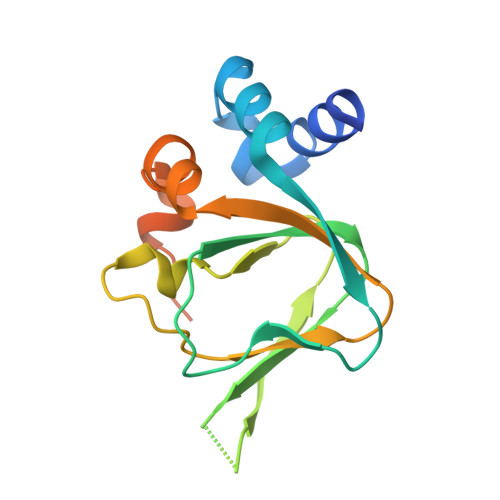
    Cyclic guanosine monophosphate (cGMP) and cyclic AMP (cAMP)-dependent protein kinases (PKG and PKA) are closely related homologs, and the cyclic nucleotide specificity of each kinase is crucial for keeping the two signaling pathways segregated, but the molecular mechanism of cyclic nucleotide selectivity is unknown. Here, we report that the PKG Iβ C-terminal cyclic nucleotide binding domain (CNB-B) is highly selective for cGMP binding, and we have solved crystal structures of CNB-B with and without bound cGMP. These structures, combined with a comprehensive mutagenic analysis, allowed us to identify Leu296 and Arg297 as key residues that mediate cGMP selectivity. In addition, by comparing the cGMP bound and unbound structures, we observed large conformational changes in the C-terminal helices in response to cGMP binding, which were stabilized by recruitment of Tyr351 as a "capping residue" for cGMP. The observed rearrangements of the C-terminal helices provide a mechanical insight into release of the catalytic domain and kinase activation.


  • Organizational Affiliation

    Verna and Marrs McLean Department of Biochemistry and Molecular Biology, Baylor College of Medicine, Houston, TX 77030, USA.


Macromolecules
Find similar proteins by:  (by identity cutoff)  |  3D Structure
Entity ID: 1
MoleculeChains Sequence LengthOrganismDetailsImage
cGMP-dependent protein kinase 1153Homo sapiensMutation(s): 0 
Gene Names: PRKG1PRKG1BPRKGR1APRKGR1B
UniProt & NIH Common Fund Data Resources
Find proteins for Q13976 (Homo sapiens)
Explore Q13976 
Go to UniProtKB:  Q13976
PHAROS:  Q13976
GTEx:  ENSG00000185532 
Entity Groups  
Sequence Clusters30% Identity50% Identity70% Identity90% Identity95% Identity100% Identity
UniProt GroupQ13976
Sequence Annotations
Expand
  • Reference Sequence
Small Molecules
Ligands 2 Unique
IDChains Name / Formula / InChI Key2D Diagram3D Interactions
PCG
Query on PCG

Download Ideal Coordinates CCD File 
Z [auth A]CYCLIC GUANOSINE MONOPHOSPHATE
C10 H12 N5 O7 P
ZOOGRGPOEVQQDX-UUOKFMHZSA-N
IOD
Query on IOD

Download Ideal Coordinates CCD File 
B [auth A]
C [auth A]
D [auth A]
E [auth A]
F [auth A]
B [auth A],
C [auth A],
D [auth A],
E [auth A],
F [auth A],
G [auth A],
H [auth A],
I [auth A],
J [auth A],
K [auth A],
L [auth A],
M [auth A],
N [auth A],
O [auth A],
P [auth A],
Q [auth A],
R [auth A],
S [auth A],
T [auth A],
U [auth A],
V [auth A],
W [auth A],
X [auth A],
Y [auth A]
IODIDE ION
I
XMBWDFGMSWQBCA-UHFFFAOYSA-M
Binding Affinity Annotations 
IDSourceBinding Affinity
PCG Binding MOAD:  4KU7 Kd: 12 (nM) from 1 assay(s)
Experimental Data & Validation

Experimental Data

  • Method: X-RAY DIFFRACTION
  • Resolution: 1.65 Å
  • R-Value Free: 0.230 
  • R-Value Work: 0.196 
  • R-Value Observed: 0.198 
  • Space Group: P 41 21 2
Unit Cell:
Length ( Å )Angle ( ˚ )
a = 47.118α = 90
b = 47.118β = 90
c = 101.799γ = 90
Software Package:
Software NamePurpose
AutoSolphasing
PHENIXrefinement
MOSFLMdata reduction
SCALAdata scaling

Structure Validation

View Full Validation Report


Ligand Structure Quality Assessment 


Entry History 

Deposition Data

Revision History  (Full details and data files)

  • Version 1.0: 2014-01-15
    Type: Initial release
  • Version 1.1: 2014-01-29
    Changes: Database references
  • Version 1.2: 2017-11-15
    Changes: Refinement description
  • Version 1.3: 2024-02-28
    Changes: Data collection, Database references, Derived calculations