


Crystal structure of the PUB domain of E3 ubiquitin ligase RNF31
Hu, J., Dong, A., Li, Y., Wernimont, A., Bountra, C., Arrowsmith, C.H., Edwards, A.M., Tong, Y.To be published.
Experimental Data Snapshot
wwPDB Validation 3D Report Full Report

Entity ID: 1 | |||||
|---|---|---|---|---|---|
| Molecule | Chains | Sequence Length | Organism | Details | Image |
| E3 ubiquitin-protein ligase RNF31 | 198 | Homo sapiens | Mutation(s): 0 Gene Names: RNF31, ZIBRA | 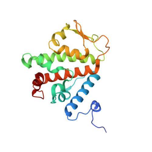 | |
UniProt & NIH Common Fund Data Resources | |||||
Find proteins for Q96EP0 (Homo sapiens) Explore Q96EP0 Go to UniProtKB: Q96EP0 | |||||
PHAROS: Q96EP0 GTEx: ENSG00000092098 | |||||
Entity Groups | |||||
| Sequence Clusters | 30% Identity50% Identity70% Identity90% Identity95% Identity100% Identity | ||||
| UniProt Group | Q96EP0 | ||||
Sequence AnnotationsExpand | |||||
| |||||
| Ligands 1 Unique | |||||
|---|---|---|---|---|---|
| ID | Chains | Name / Formula / InChI Key | 2D Diagram | 3D Interactions | |
| UNX Query on UNX | C [auth A] D [auth A] E [auth A] F [auth A] G [auth A] | UNKNOWN ATOM OR ION X |  | ||
| Length ( Å ) | Angle ( ˚ ) |
|---|---|
| a = 256.488 | α = 90 |
| b = 256.488 | β = 90 |
| c = 256.488 | γ = 90 |
| Software Name | Purpose |
|---|---|
| SOLVE | phasing |
| REFMAC | refinement |
| PDB_EXTRACT | data extraction |
| SBC-Collect | data collection |
| HKL-3000 | data reduction |
| HKL-3000 | data scaling |
RCSB PDB (citation) is hosted by
RCSB PDB is a member of the

