4J6S

14-3-3gamma complexed with the N-terminal sequence of tyrosine hydroxylase (residues 1-43)


Experimental Data Snapshot

  • Method: X-RAY DIFFRACTION
  • Resolution: 3.08 Å
  • R-Value Free: 0.254 
  • R-Value Work: 0.210 
  • R-Value Observed: 0.212 

wwPDB Validation   3D Report Full Report


This is version 1.3 of the entry. See complete history


Literature

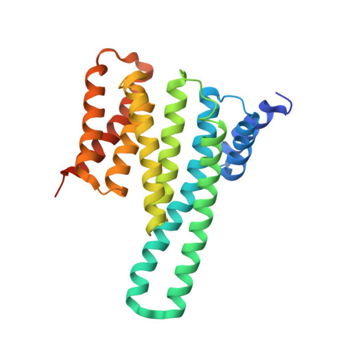
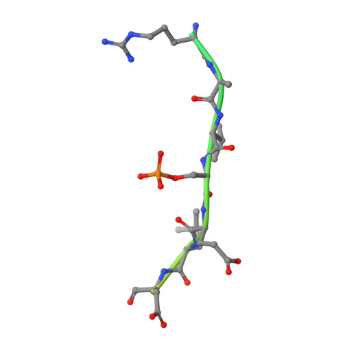
The N-terminal sequence of tyrosine hydroxylase is a conformationally versatile motif that binds 14-3-3 proteins and membranes.

Skjevik, A.A.Mileni, M.Baumann, A.Halskau, O.Teigen, K.Stevens, R.C.Martinez, A.

(2014) J Mol Biol 426: 150-168

  • DOI: https://doi.org/10.1016/j.jmb.2013.09.012
  • Primary Citation of Related Structures:  
    4J6S

  • PubMed Abstract: 

    Tyrosine hydroxylase (TH) catalyzes the rate-limiting step in the synthesis of catecholamine neurotransmitters, and a reduction in TH activity is associated with several neurological diseases. Human TH is regulated, among other mechanisms, by Ser19-phosphorylation-dependent interaction with 14-3-3 proteins. The N-terminal sequence (residues 1-43), which corresponds to an extension to the TH regulatory domain, also interacts with negatively charged membranes. By using X-ray crystallography together with molecular dynamics simulations and structural bioinformatics analysis, we have probed the conformations of the Ser19-phosphorylated N-terminal peptide [THp-(1-43)] bound to 14-3-3γ, free in solution and bound to a phospholipid bilayer, and of the unphosphorylated peptide TH-(1-43) both free and bilayer bound. As seen in the crystal structure of THp-(1-43) complexed with 14-3-3γ, the region surrounding pSer19 adopts an extended conformation in the bound state, whereas THp-(1-43) adopts a bent conformation when free in solution, with higher content of secondary structure and higher number of internal hydrogen bonds. TH-(1-43) in solution presents the highest mobility and least defined structure of all forms studied, and it shows an energetically more favorable interaction with membranes relative to THp-(1-43). Cationic residues, notably Arg15 and Arg16, which are the recognition sites of the kinases phosphorylating at Ser19, are also contributing to the interaction with the membrane. Our results reveal the structural flexibility of this region of TH, in accordance with the functional versatility and conformational adaptation to different partners. Furthermore, this structural information has potential relevance for the development of therapeutics for neurodegenerative disorders, through modulation of TH-partner interactions.


  • Organizational Affiliation

    Department of Biomedicine, University of Bergen, Jonas Lies vei 91, 5009 Bergen, Norway.


Macromolecules
Find similar proteins by:  (by identity cutoff)  |  3D Structure
Entity ID: 1
MoleculeChains Sequence LengthOrganismDetailsImage
14-3-3 protein gamma
A, B, C, D
255Homo sapiensMutation(s): 0 
Gene Names: YWHAG
UniProt & NIH Common Fund Data Resources
Find proteins for P61981 (Homo sapiens)
Explore P61981 
Go to UniProtKB:  P61981
PHAROS:  P61981
GTEx:  ENSG00000170027 
Entity Groups  
Sequence Clusters30% Identity50% Identity70% Identity90% Identity95% Identity100% Identity
UniProt GroupP61981
Sequence Annotations
Expand
  • Reference Sequence
Find similar proteins by:  (by identity cutoff)  |  3D Structure
Entity ID: 2
MoleculeChains Sequence LengthOrganismDetailsImage
N-terminal motif of tyrosine hydroxylase
E, F, G, H
43Homo sapiensMutation(s): 0 
EC: 1.14.16.2
UniProt & NIH Common Fund Data Resources
Find proteins for P07101 (Homo sapiens)
Explore P07101 
Go to UniProtKB:  P07101
PHAROS:  P07101
GTEx:  ENSG00000180176 
Entity Groups  
Sequence Clusters30% Identity50% Identity70% Identity90% Identity95% Identity100% Identity
UniProt GroupP07101
Sequence Annotations
Expand
  • Reference Sequence
Small Molecules
Modified Residues  1 Unique
IDChains TypeFormula2D DiagramParent
SEP
Query on SEP
E, F, G, H
L-PEPTIDE LINKINGC3 H8 N O6 PSER
Experimental Data & Validation

Experimental Data

  • Method: X-RAY DIFFRACTION
  • Resolution: 3.08 Å
  • R-Value Free: 0.254 
  • R-Value Work: 0.210 
  • R-Value Observed: 0.212 
  • Space Group: P 21 21 21
Unit Cell:
Length ( Å )Angle ( ˚ )
a = 84.19α = 90
b = 115.12β = 90
c = 136.9γ = 90
Software Package:
Software NamePurpose
XDSdata scaling
PHASERphasing
REFMACrefinement
XDSdata reduction

Structure Validation

View Full Validation Report


Entry History 

Deposition Data

Revision History  (Full details and data files)

  • Version 1.0: 2013-10-02
    Type: Initial release
  • Version 1.1: 2014-01-08
    Changes: Database references
  • Version 1.2: 2021-04-21
    Changes: Database references, Derived calculations
  • Version 1.3: 2023-09-20
    Changes: Data collection, Database references, Refinement description