4IQ6

Gsk-3beta with inhibitor 6-chloro-N-cyclohexyl-4-(1H-pyrrolo[2,3-b]pyridin-3-yl)pyridin-2-amine


Experimental Data Snapshot

  • Method: X-RAY DIFFRACTION
  • Resolution: 3.12 Å
  • R-Value Free: 0.227 
  • R-Value Work: 0.186 
  • R-Value Observed: 0.188 

wwPDB Validation   3D Report Full Report


Ligand Structure Quality Assessment 


This is version 1.3 of the entry. See complete history


Literature

Azaindole-Based Inhibitors of Cdc7 Kinase: Impact of the Pre-DFG Residue, Val 195.

Tong, Y.Stewart, K.D.Florjancic, A.S.Harlan, J.E.Merta, P.J.Przytulinska, M.Soni, N.Swinger, K.K.Zhu, H.Johnson, E.F.Shoemaker, A.R.Penning, T.D.

(2013) ACS Med Chem Lett 4: 211-215

  • DOI: https://doi.org/10.1021/ml300348c
  • Primary Citation of Related Structures:  
    4IQ6

  • PubMed Abstract: 

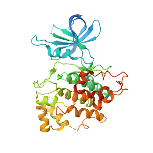
    To investigate the role played by the unique pre-DFG residue Val 195 of Cdc7 kinase on the potency of azaindole-chloropyridines (1), a series of novel analogues with various chloro replacements were synthesized and evaluated for their inhibitory activity against Cdc7. X-ray cocrystallization using a surrogate protein, GSK3β, and modeling studies confirmed the azaindole motif as the hinge binder. Weaker hydrophobic interactions with Met 134 and Val 195 by certain chloro replacements (e.g., H, methyl) led to reduced Cdc7 inhibition. Meanwhile, data from other replacements (e.g., F, O) indicated that loss of such hydrophobic interaction could be compensated by enhanced hydrogen bonding to Lys 90. Our findings not only provide an in-depth understanding of the pre-DFG residue as another viable position impacting kinase inhibition, they also expand the existing knowledge of ligand-Cdc7 binding.


  • Organizational Affiliation

    Cancer Research, Structural Biology, Lead Discovery, and Protein Biochemistry, Global Pharmaceutical Research and Development, Abbott Laboratories , 100 Abbott Park Road, Abbott Park, Illinois 60064, United States.


Macromolecules
Find similar proteins by:  (by identity cutoff)  |  3D Structure
Entity ID: 1
MoleculeChains Sequence LengthOrganismDetailsImage
Glycogen synthase kinase-3 beta
A, B
426Homo sapiensMutation(s): 0 
Gene Names: GSK3B
EC: 2.7.11.26 (PDB Primary Data), 2.7.11.1 (PDB Primary Data)
UniProt & NIH Common Fund Data Resources
Find proteins for P49841 (Homo sapiens)
Explore P49841 
Go to UniProtKB:  P49841
PHAROS:  P49841
GTEx:  ENSG00000082701 
Entity Groups  
Sequence Clusters30% Identity50% Identity70% Identity90% Identity95% Identity100% Identity
UniProt GroupP49841
Sequence Annotations
Expand
  • Reference Sequence
Small Molecules
Ligands 1 Unique
IDChains Name / Formula / InChI Key2D Diagram3D Interactions
IQ6
Query on IQ6

Download Ideal Coordinates CCD File 
C [auth A],
D [auth B]
6-chloro-N-cyclohexyl-4-(1H-pyrrolo[2,3-b]pyridin-3-yl)pyridin-2-amine
C18 H19 Cl N4
JKTMEYXRCJFWAI-UHFFFAOYSA-N
Binding Affinity Annotations 
IDSourceBinding Affinity
IQ6 PDBBind:  4IQ6 Ki: 58 (nM) from 1 assay(s)
BindingDB:  4IQ6 Ki: 58 (nM) from 1 assay(s)
Experimental Data & Validation

Experimental Data

  • Method: X-RAY DIFFRACTION
  • Resolution: 3.12 Å
  • R-Value Free: 0.227 
  • R-Value Work: 0.186 
  • R-Value Observed: 0.188 
  • Space Group: P 21 21 21
Unit Cell:
Length ( Å )Angle ( ˚ )
a = 82.835α = 90
b = 84.991β = 90
c = 178.058γ = 90
Software Package:
Software NamePurpose
JDirectordata collection
MOLREPphasing
BUSTERrefinement
XDSdata reduction
autoPROCdata scaling

Structure Validation

View Full Validation Report


Ligand Structure Quality Assessment 


Entry History 

Revision History  (Full details and data files)

  • Version 1.0: 2013-04-24
    Type: Initial release
  • Version 1.1: 2014-09-24
    Changes: Database references
  • Version 1.2: 2017-11-15
    Changes: Refinement description
  • Version 1.3: 2024-02-28
    Changes: Data collection, Database references, Derived calculations, Refinement description