4IF5

Structure of human Mec17


Experimental Data Snapshot

  • Method: X-RAY DIFFRACTION
  • Resolution: 1.70 Å
  • R-Value Free: 0.214 
  • R-Value Work: 0.178 
  • R-Value Observed: 0.182 

wwPDB Validation   3D Report Full Report


Ligand Structure Quality Assessment 


This is version 1.1 of the entry. See complete history


Literature

Structural and Functional Characterization of the alpha-Tubulin Acetyltransferase MEC-17.

Davenport, A.M.Collins, L.N.Chiu, H.Minor, P.J.Sternberg, P.W.Hoelz, A.

(2014) J Mol Biol 426: 2605-2616

  • DOI: https://doi.org/10.1016/j.jmb.2014.05.009
  • Primary Citation of Related Structures:  
    4IF5

  • PubMed Abstract: 

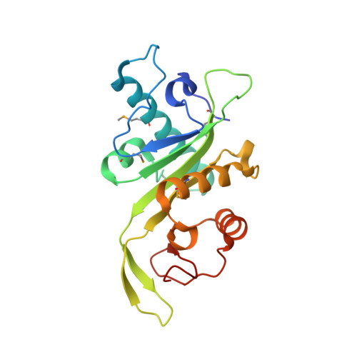
    Tubulin protomers undergo an extensive array of post-translational modifications to tailor microtubules to specific tasks. One such modification, the acetylation of lysine 40 of α-tubulin, located in the lumen of microtubules, is associated with stable, long-living microtubule structures. MEC-17 was recently identified as the acetyltransferase that mediates this event. We have determined the crystal structure of the catalytic core of human MEC-17 in complex with its cofactor acetyl-CoA at 1.7Å resolution. The structure reveals that the MEC-17 core adopts a canonical Gcn5-related N-acetyltransferase (GNAT) fold that is decorated with extensive surface loops. An enzymatic analysis of 33 MEC-17 surface mutants identifies hot-spot residues for catalysis and substrate recognition. A large, evolutionarily conserved hydrophobic surface patch that is critical for enzymatic activity is identified, suggesting that specificity is achieved by interactions with the α-tubulin substrate that extend outside of the modified surface loop. An analysis of MEC-17 mutants in Caenorhabditis elegans shows that enzymatic activity is dispensable for touch sensitivity.


  • Organizational Affiliation

    Division of Chemistry and Chemical Engineering, California Institute of Technology, 1200 East California Boulevard, Pasadena, CA 91125, USA.


Macromolecules
Find similar proteins by:  (by identity cutoff)  |  3D Structure
Entity ID: 1
MoleculeChains Sequence LengthOrganismDetailsImage
Alpha-tubulin N-acetyltransferase196Homo sapiensMutation(s): 0 
Gene Names: ATAT1C6orf134MEC17Nbla00487
EC: 2.3.1.108
UniProt & NIH Common Fund Data Resources
Find proteins for Q5SQI0 (Homo sapiens)
Explore Q5SQI0 
Go to UniProtKB:  Q5SQI0
PHAROS:  Q5SQI0
GTEx:  ENSG00000137343 
Entity Groups  
Sequence Clusters30% Identity50% Identity70% Identity90% Identity95% Identity100% Identity
UniProt GroupQ5SQI0
Sequence Annotations
Expand
  • Reference Sequence
Small Molecules
Ligands 2 Unique
IDChains Name / Formula / InChI Key2D Diagram3D Interactions
ACO
Query on ACO

Download Ideal Coordinates CCD File 
B [auth A]ACETYL COENZYME *A
C23 H38 N7 O17 P3 S
ZSLZBFCDCINBPY-ZSJPKINUSA-N
CL
Query on CL

Download Ideal Coordinates CCD File 
C [auth A]CHLORIDE ION
Cl
VEXZGXHMUGYJMC-UHFFFAOYSA-M
Modified Residues  1 Unique
IDChains TypeFormula2D DiagramParent
MSE
Query on MSE
A
L-PEPTIDE LINKINGC5 H11 N O2 SeMET
Experimental Data & Validation

Experimental Data

  • Method: X-RAY DIFFRACTION
  • Resolution: 1.70 Å
  • R-Value Free: 0.214 
  • R-Value Work: 0.178 
  • R-Value Observed: 0.182 
  • Space Group: P 21 21 2
Unit Cell:
Length ( Å )Angle ( ˚ )
a = 41.747α = 90
b = 122.297β = 90
c = 37.541γ = 90
Software Package:
Software NamePurpose
PHENIXrefinement
PDB_EXTRACTdata extraction
HKL-2000data collection
HKL-2000data reduction
HKL-2000data scaling
SHARPphasing

Structure Validation

View Full Validation Report


Ligand Structure Quality Assessment 


Entry History 

Deposition Data

Revision History  (Full details and data files)

  • Version 1.0: 2014-05-28
    Type: Initial release
  • Version 1.1: 2014-07-02
    Changes: Database references