4IC3

Crystal structure of the F495L mutant XIAP RING domain


Experimental Data Snapshot

  • Method: X-RAY DIFFRACTION
  • Resolution: 1.78 Å
  • R-Value Free: 0.222 
  • R-Value Work: 0.176 
  • R-Value Observed: 0.178 

wwPDB Validation   3D Report Full Report


This is version 1.2 of the entry. See complete history


Literature

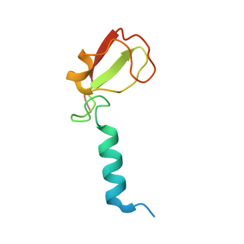
Regulation of ubiquitin transfer by XIAP, a dimeric RING E3 ligase

Nakatani, Y.Kleffmann, T.Linke, K.Condon, S.M.Hinds, M.G.Day, C.L.

(2013) Biochem J 450: 629-638

  • DOI: https://doi.org/10.1042/BJ20121702
  • Primary Citation of Related Structures:  
    4IC2, 4IC3

  • PubMed Abstract: 

    RING domains of E3 ligases promote transfer of Ub (ubiquitin) from the E2~Ub conjugate to target proteins. In many cases interaction of the E2~Ub conjugate with the RING domain requires its prior dimerization. Using cross-linking experiments we show that E2 conjugated ubiquitin contacts the RING homodimer interface of the IAP (inhibitor of apoptosis) proteins, XIAP (X-linked IAP) and cIAP (cellular IAP) 2. Structural and biochemical analysis of the XIAP RING dimer shows that an aromatic residue at the dimer interface is required for E2~Ub binding and Ub transfer. Mutation of the aromatic residue abolishes Ub transfer, but not interaction with Ub. This indicates that nuleophilic attack on the thioester bond depends on precise contacts between Ub and the RING domain. RING dimerization is a critical activating step for the cIAP proteins; however, our analysis shows that the RING domain of XIAP forms a stable dimer and its E3 ligase activity does not require an activation step.


  • Organizational Affiliation

    Biochemistry Department, Otago School of Medical Sciences, University of Otago, Dunedin 9054, New Zealand.


Macromolecules
Find similar proteins by:  (by identity cutoff)  |  3D Structure
Entity ID: 1
MoleculeChains Sequence LengthOrganismDetailsImage
E3 ubiquitin-protein ligase XIAP
A, B
74Homo sapiensMutation(s): 1 
Gene Names: API3BIR4IAP3XIAP
EC: 6.3.2
UniProt & NIH Common Fund Data Resources
Find proteins for P98170 (Homo sapiens)
Explore P98170 
Go to UniProtKB:  P98170
PHAROS:  P98170
GTEx:  ENSG00000101966 
Entity Groups  
Sequence Clusters30% Identity50% Identity70% Identity90% Identity95% Identity100% Identity
UniProt GroupP98170
Sequence Annotations
Expand
  • Reference Sequence
Experimental Data & Validation

Experimental Data

  • Method: X-RAY DIFFRACTION
  • Resolution: 1.78 Å
  • R-Value Free: 0.222 
  • R-Value Work: 0.176 
  • R-Value Observed: 0.178 
  • Space Group: P 1 21 1
Unit Cell:
Length ( Å )Angle ( ˚ )
a = 35.735α = 90
b = 48.471β = 97.47
c = 39.967γ = 90
Software Package:
Software NamePurpose
Blu-Icedata collection
PHASERphasing
PHENIXrefinement
XDSdata reduction
SCALAdata scaling

Structure Validation

View Full Validation Report


Entry History 

Deposition Data

Revision History  (Full details and data files)

  • Version 1.0: 2012-12-19
    Type: Initial release
  • Version 1.1: 2013-07-10
    Changes: Database references
  • Version 1.2: 2023-11-08
    Changes: Data collection, Database references, Derived calculations, Refinement description