4I22

Structure of the monomeric (V948R)gefitinib/erlotinib resistant double mutant (L858R+T790M) EGFR kinase domain co-crystallized with gefitinib


Experimental Data Snapshot

  • Method: X-RAY DIFFRACTION
  • Resolution: 1.71 Å
  • R-Value Free: 0.224 
  • R-Value Work: 0.197 
  • R-Value Observed: 0.198 

wwPDB Validation   3D Report Full Report


Ligand Structure Quality Assessment 


This is version 1.4 of the entry. See complete history


Literature

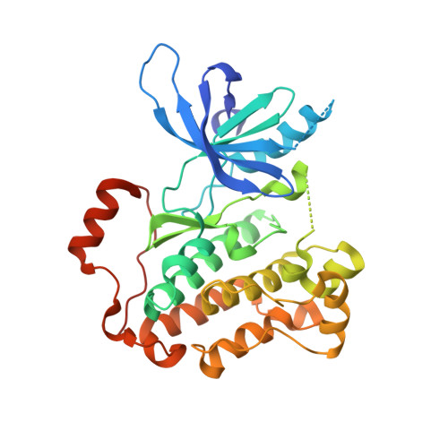
Insights into the Aberrant Activity of Mutant EGFR Kinase Domain and Drug Recognition.

Gajiwala, K.S.Feng, J.Ferre, R.Ryan, K.Brodsky, O.Weinrich, S.Kath, J.C.Stewart, A.

(2013) Structure 21: 209-219

  • DOI: https://doi.org/10.1016/j.str.2012.11.014
  • Primary Citation of Related Structures:  
    4I1Z, 4I20, 4I21, 4I22, 4I23, 4I24

  • PubMed Abstract: 

    The oncogenicity of the L858R mutant form of the epidermal growth factor receptor (EGFR) in non-small-cell lung cancer is thought to be due to the constitutive activation of its kinase domain. The selectivity of the marketed drugs gefitinib and erlotinib for L858R mutant is attributed to their specific recognition of the active kinase and to weaker ATP binding by L858R EGFR. We present crystal structures showing that neither L858R nor the drug-resistant L858R+T790M EGFR kinase domain is in the constitutively active conformation. Additional co-crystal structures show that gefitinib and dacomitinib, an irreversible anilinoquinazoline derivative currently in clinical development, may not be conformation specific for the active state of the enzyme. Structural data further reveal the potential mode of recognition of one of the autophosphorylation sites in the C-terminal tail, Tyr-1016, by the kinase domain. Biochemical and biophysical evidence suggest that the oncogenic mutations impact the conformational dynamics of the enzyme.


  • Organizational Affiliation

    Cancer Structural Biology, Oncology Medicinal Chemistry, Pfizer Worldwide Research and Development, 10770 Science Center Drive, San Diego, CA 92121, USA. ketan.gajiwala@pfizer.com


Macromolecules
Find similar proteins by:  (by identity cutoff)  |  3D Structure
Entity ID: 1
MoleculeChains Sequence LengthOrganismDetailsImage
Epidermal growth factor receptor329Homo sapiensMutation(s): 3 
Gene Names: EGFRERBBERBB1HER1
EC: 2.7.10.1
UniProt & NIH Common Fund Data Resources
Find proteins for P00533 (Homo sapiens)
Explore P00533 
Go to UniProtKB:  P00533
PHAROS:  P00533
GTEx:  ENSG00000146648 
Entity Groups  
Sequence Clusters30% Identity50% Identity70% Identity90% Identity95% Identity100% Identity
UniProt GroupP00533
Sequence Annotations
Expand
  • Reference Sequence
Experimental Data & Validation

Experimental Data

  • Method: X-RAY DIFFRACTION
  • Resolution: 1.71 Å
  • R-Value Free: 0.224 
  • R-Value Work: 0.197 
  • R-Value Observed: 0.198 
  • Space Group: P 1 2 1
Unit Cell:
Length ( Å )Angle ( ˚ )
a = 76.519α = 90
b = 35.486β = 112.89
c = 77.781γ = 90
Software Package:
Software NamePurpose
JDirectordata collection
CNXrefinement
autoPROCdata scaling
SCALAdata scaling
CNXphasing

Structure Validation

View Full Validation Report


Ligand Structure Quality Assessment 


Entry History 

Deposition Data

Revision History  (Full details and data files)

  • Version 1.0: 2013-01-16
    Type: Initial release
  • Version 1.1: 2013-02-27
    Changes: Database references
  • Version 1.2: 2014-10-15
    Changes: Non-polymer description
  • Version 1.3: 2017-11-15
    Changes: Refinement description
  • Version 1.4: 2024-02-28
    Changes: Data collection, Database references, Derived calculations, Refinement description