4HTP

Crystal structure of the DBD domain of human DNA ligase IV bound to Artemis peptide


Experimental Data Snapshot

  • Method: X-RAY DIFFRACTION
  • Resolution: 2.25 Å
  • R-Value Free: 0.249 
  • R-Value Work: 0.196 
  • R-Value Observed: 0.199 

wwPDB Validation   3D Report Full Report


This is version 1.2 of the entry. See complete history


Literature

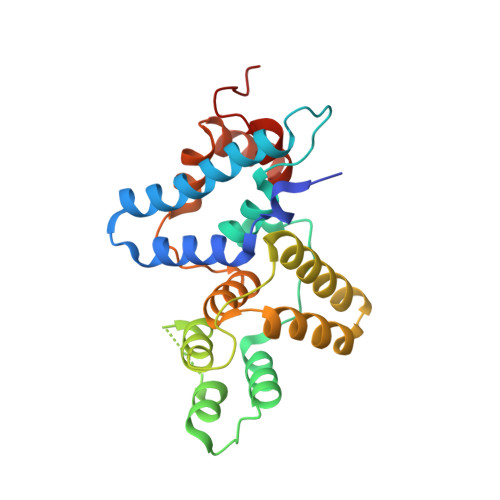
Structural Basis of DNA Ligase IV-Artemis Interaction in Nonhomologous End-Joining.

De Ioannes, P.Malu, S.Cortes, P.Aggarwal, A.K.

(2012) Cell Rep 2: 1505-1512

  • DOI: https://doi.org/10.1016/j.celrep.2012.11.004
  • Primary Citation of Related Structures:  
    4HTO, 4HTP

  • PubMed Abstract: 

    DNA ligase IV (LigIV) and Artemis are central components of the nonhomologous end-joining (NHEJ) machinery that is required for V(D)J recombination and the maintenance of genomic integrity in mammalian cells. We report here crystal structures of the LigIV DNA binding domain (DBD) in both its apo form and in complex with a peptide derived from the Artemis C-terminal region. We show that LigIV interacts with Artemis through an extended hydrophobic surface. In particular, we find that the helix α2 in LigIV-DBD is longer than in other mammalian ligases and presents residues that specifically interact with the Artemis peptide, which adopts a partially helical conformation on binding. Mutations of key residues on the LigIV-DBD hydrophobic surface abolish the interaction. Together, our results provide structural insights into the specificity of the LigIV-Artemis interaction and how the enzymatic activities of the two proteins may be coordinated during NHEJ.


  • Organizational Affiliation

    Department of Structural and Chemical Biology, Mount Sinai School of Medicine, New York, NY 10029, USA.


Macromolecules
Find similar proteins by:  (by identity cutoff)  |  3D Structure
Entity ID: 1
MoleculeChains Sequence LengthOrganismDetailsImage
DNA ligase 4
A, B
240Homo sapiensMutation(s): 0 
Gene Names: LIG4
EC: 6.5.1.1
UniProt & NIH Common Fund Data Resources
Find proteins for P49917 (Homo sapiens)
Explore P49917 
Go to UniProtKB:  P49917
PHAROS:  P49917
GTEx:  ENSG00000174405 
Entity Groups  
Sequence Clusters30% Identity50% Identity70% Identity90% Identity95% Identity100% Identity
UniProt GroupP49917
Sequence Annotations
Expand
  • Reference Sequence

Find similar proteins by:  Sequence   |   3D Structure  

Entity ID: 2
MoleculeChains Sequence LengthOrganismDetailsImage
Protein artemisC,
D [auth E]
11Homo sapiensMutation(s): 0 
EC: 3.1
UniProt & NIH Common Fund Data Resources
Find proteins for Q96SD1 (Homo sapiens)
Explore Q96SD1 
Go to UniProtKB:  Q96SD1
PHAROS:  Q96SD1
GTEx:  ENSG00000152457 
Entity Groups  
Sequence Clusters30% Identity50% Identity70% Identity90% Identity95% Identity100% Identity
UniProt GroupQ96SD1
Sequence Annotations
Expand
  • Reference Sequence
Experimental Data & Validation

Experimental Data

  • Method: X-RAY DIFFRACTION
  • Resolution: 2.25 Å
  • R-Value Free: 0.249 
  • R-Value Work: 0.196 
  • R-Value Observed: 0.199 
  • Space Group: P 21 21 21
Unit Cell:
Length ( Å )Angle ( ˚ )
a = 61.21α = 90
b = 72.83β = 90
c = 158.05γ = 90
Software Package:
Software NamePurpose
HKL-2000data collection
PHASERphasing
PHENIXrefinement
HKL-2000data reduction
HKL-2000data scaling

Structure Validation

View Full Validation Report


Entry History 

Deposition Data

Revision History  (Full details and data files)

  • Version 1.0: 2012-12-26
    Type: Initial release
  • Version 1.1: 2013-01-16
    Changes: Database references
  • Version 1.2: 2023-09-20
    Changes: Data collection, Database references, Refinement description