4HQR

Crystal Structure of mutant form of Caspase-7


Experimental Data Snapshot

  • Method: X-RAY DIFFRACTION
  • Resolution: 3.00 Å
  • R-Value Free: 0.253 
  • R-Value Work: 0.218 
  • R-Value Observed: 0.221 

wwPDB Validation   3D Report Full Report


This is version 1.2 of the entry. See complete history


Literature

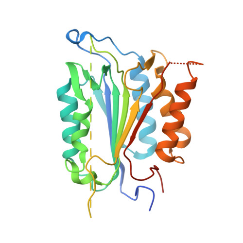
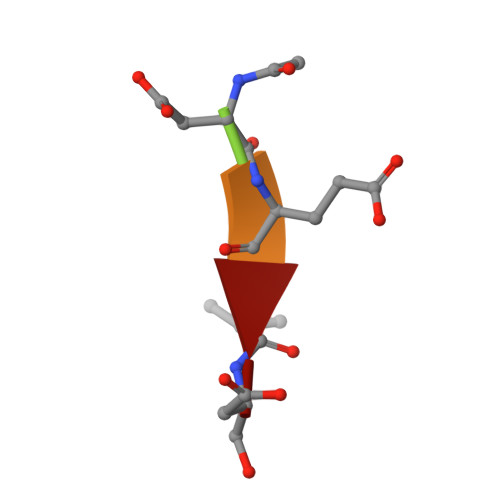
Structural asymmetry of procaspase-7 bound to a specific inhibitor

Kang, H.J.Lee, Y.M.Bae, K.H.Kim, S.J.Chung, S.J.

(2013) Acta Crystallogr D Biol Crystallogr 69: 1514-1521

  • DOI: https://doi.org/10.1107/S0907444913010196
  • Primary Citation of Related Structures:  
    4HQ0, 4HQR

  • PubMed Abstract: 

    Caspase-7 is expressed as a proenzyme and is activated by initiator caspases upon the transmission of cell-death signals. Despite extensive structural and biochemical analyses, many questions regarding the mechanism of caspase-7 activation remain unanswered. Caspase-7 is auto-activated during overexpression in Escherichia coli, even in the absence of initiator caspases, indicating that procaspase-7 has intrinsic catalytic activity. When variants of procaspase-7 with altered L2 loops were prepared, a variant with six inserted amino acids showed meaningful catalytic activity which was inhibited by Ac-DEVD-CHO. The kinetic constants of the procaspase-7 variant were determined and its three-dimensional structure was solved with and without bound inhibitor. The homodimeric procaspase-7 bound to the inhibitor revealed an asymmetry. One monomer formed a complete active site bound to the inhibitor in collaboration with the L2 loop from the other monomer, whereas the other monomer had an incomplete active site despite the bound inhibitor. Consequently, the two L2 loops in homodimeric procaspase-7 served as inherent L2 and L2' loops forming one complete active site. These data represent the first three-dimensional structure of a procaspase-7 variant bound to a specific inhibitor, Ac-DEVD-CHO, and provide insight into the folding mechanism during caspase-7 activation and the basal activity level of procaspase-7.


  • Organizational Affiliation

    Department of Chemistry, College of Natural Science, Dongguk University, 26 Pil-dong 3-ga, Jung-gu, Seoul 100-715, Republic of Korea.


Macromolecules
Find similar proteins by:  (by identity cutoff)  |  3D Structure
Entity ID: 1
MoleculeChains Sequence LengthOrganismDetailsImage
Caspase-7
A, B
272Homo sapiensMutation(s): 1 
UniProt & NIH Common Fund Data Resources
Find proteins for P55210 (Homo sapiens)
Explore P55210 
Go to UniProtKB:  P55210
PHAROS:  P55210
GTEx:  ENSG00000165806 
Entity Groups  
Sequence Clusters30% Identity50% Identity70% Identity90% Identity95% Identity100% Identity
UniProt GroupP55210
Sequence Annotations
Expand
  • Reference Sequence

Find similar proteins by:  Sequence   |   3D Structure  

Entity ID: 2
MoleculeChains Sequence LengthOrganismDetailsImage
Ac-Asp-Glu-Val-Asp-AldehydeC [auth E],
D [auth F]
5N/AMutation(s): 0 
Sequence Annotations
Expand
  • Reference Sequence
Biologically Interesting Molecules (External Reference) 1 Unique
Experimental Data & Validation

Experimental Data

  • Method: X-RAY DIFFRACTION
  • Resolution: 3.00 Å
  • R-Value Free: 0.253 
  • R-Value Work: 0.218 
  • R-Value Observed: 0.221 
  • Space Group: P 32 2 1
Unit Cell:
Length ( Å )Angle ( ˚ )
a = 90.48α = 90
b = 90.48β = 90
c = 185.05γ = 120
Software Package:
Software NamePurpose
ADSCdata collection
PHASERphasing
CNSrefinement
MOSFLMdata reduction
SCALAdata scaling

Structure Validation

View Full Validation Report


Entry History 

Deposition Data

Revision History  (Full details and data files)

  • Version 1.0: 2013-09-11
    Type: Initial release
  • Version 1.1: 2023-11-08
    Changes: Data collection, Database references, Derived calculations, Refinement description
  • Version 1.2: 2023-12-06
    Changes: Data collection