4H0W

Bismuth bound human serum transferrin


Experimental Data Snapshot

  • Method: X-RAY DIFFRACTION
  • Resolution: 2.40 Å
  • R-Value Free: 0.256 
  • R-Value Work: 0.194 
  • R-Value Observed: 0.197 

wwPDB Validation   3D Report Full Report


Ligand Structure Quality Assessment 


This is version 2.0 of the entry. See complete history


Literature

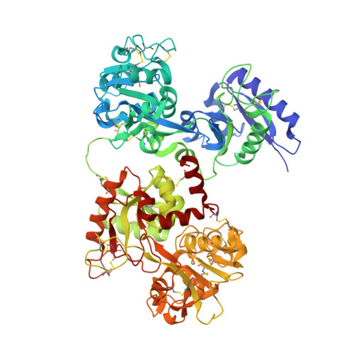
Iron and bismuth bound human serum transferrin reveals a partially-opened conformation in the N-lobe.

Yang, N.Zhang, H.Wang, M.Hao, Q.Sun, H.

(2012) Sci Rep 2: 999-999

  • DOI: https://doi.org/10.1038/srep00999
  • Primary Citation of Related Structures:  
    3QYT, 4H0W

  • PubMed Abstract: 

    Human serum transferrin (hTF) binds Fe(III) tightly but reversibly, and delivers it to cells via a receptor-mediated endocytosis process. The metal-binding and release result in significant conformational changes of the protein. Here, we report the crystal structures of diferric-hTF (Fe(N)Fe(C)-hTF) and bismuth-bound hTF (Bi(N)Fe(C)-hTF) at 2.8 and 2.4 Å resolutions respectively. Notably, the N-lobes of both structures exhibit unique "partially-opened" conformations between those of the apo-hTF and holo-hTF. Fe(III) and Bi(III) in the N-lobe coordinate to, besides anions, only two (Tyr95 and Tyr188) and one (Tyr188) tyrosine residues, respectively, in contrast to four residues in the holo-hTF. The C-lobe of both structures are fully closed with iron coordinating to four residues and a carbonate. The structures of hTF observed here represent key conformers captured in the dynamic nature of the transferrin family proteins and provide a structural basis for understanding the mechanism of metal uptake and release in transferrin families.


  • Organizational Affiliation

    Department of Chemistry, the University of Hong Kong, Pokfulam Road, Hong Kong, PR China.


Macromolecules
Find similar proteins by:  (by identity cutoff)  |  3D Structure
Entity ID: 1
MoleculeChains Sequence LengthOrganismDetailsImage
Serotransferrin679Homo sapiensMutation(s): 0 
UniProt & NIH Common Fund Data Resources
Find proteins for P02787 (Homo sapiens)
Explore P02787 
Go to UniProtKB:  P02787
PHAROS:  P02787
GTEx:  ENSG00000091513 
Entity Groups  
Sequence Clusters30% Identity50% Identity70% Identity90% Identity95% Identity100% Identity
UniProt GroupP02787
Sequence Annotations
Expand
  • Reference Sequence
Small Molecules
Ligands 5 Unique
IDChains Name / Formula / InChI Key2D Diagram3D Interactions
NAG
Query on NAG

Download Ideal Coordinates CCD File 
F [auth A]2-acetamido-2-deoxy-beta-D-glucopyranose
C8 H15 N O6
OVRNDRQMDRJTHS-FMDGEEDCSA-N
BS3
Query on BS3

Download Ideal Coordinates CCD File 
D [auth A]Bismuth(III) ION
Bi
JCXGWMGPZLAOME-UHFFFAOYSA-N
NTA
Query on NTA

Download Ideal Coordinates CCD File 
G [auth A]NITRILOTRIACETIC ACID
C6 H9 N O6
MGFYIUFZLHCRTH-UHFFFAOYSA-N
CO3
Query on CO3

Download Ideal Coordinates CCD File 
C [auth A],
E [auth A]
CARBONATE ION
C O3
BVKZGUZCCUSVTD-UHFFFAOYSA-L
FE
Query on FE

Download Ideal Coordinates CCD File 
B [auth A]FE (III) ION
Fe
VTLYFUHAOXGGBS-UHFFFAOYSA-N
Experimental Data & Validation

Experimental Data

  • Method: X-RAY DIFFRACTION
  • Resolution: 2.40 Å
  • R-Value Free: 0.256 
  • R-Value Work: 0.194 
  • R-Value Observed: 0.197 
  • Space Group: P 21 21 21
Unit Cell:
Length ( Å )Angle ( ˚ )
a = 73.907α = 90
b = 90.165β = 90
c = 111.032γ = 90
Software Package:
Software NamePurpose
Blu-Icedata collection
PHASERphasing
REFMACrefinement
HKL-2000data reduction
HKL-2000data scaling

Structure Validation

View Full Validation Report


Ligand Structure Quality Assessment 


Entry History 

Deposition Data

Revision History  (Full details and data files)

  • Version 1.0: 2012-12-26
    Type: Initial release
  • Version 1.1: 2013-01-16
    Changes: Database references
  • Version 1.2: 2020-07-29
    Type: Remediation
    Reason: Carbohydrate remediation
    Changes: Data collection, Derived calculations, Structure summary
  • Version 1.3: 2023-09-13
    Changes: Data collection, Database references, Refinement description, Structure summary
  • Version 2.0: 2023-11-15
    Changes: Atomic model, Data collection, Derived calculations