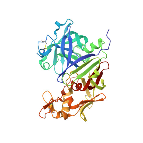
4GJ8

Crystal structure of renin in complex with PKF909-724 (compound 3)


Experimental Data Snapshot

  • Method: X-RAY DIFFRACTION
  • Resolution: 2.50 Å
  • R-Value Free: 0.217 
  • R-Value Work: 0.169 
  • R-Value Observed: 0.174 

wwPDB Validation   3D Report Full Report


Ligand Structure Quality Assessment 


This is version 1.3 of the entry. See complete history


Literature

A novel class of oral direct Renin inhibitors: highly potent 3,5-disubstituted piperidines bearing a tricyclic p3-p1 pharmacophore.

Ostermann, N.Ruedisser, S.Ehrhardt, C.Breitenstein, W.Marzinzik, A.Jacoby, E.Vangrevelinghe, E.Ottl, J.Klumpp, M.Hartwieg, J.C.Cumin, F.Hassiepen, U.Trappe, J.Sedrani, R.Geisse, S.Gerhartz, B.Richert, P.Francotte, E.Wagner, T.Kromer, M.Kosaka, T.Webb, R.L.Rigel, D.F.Maibaum, J.Baeschlin, D.K.

(2013) J Med Chem 56: 2196-2206

  • DOI: https://doi.org/10.1021/jm301706j
  • Primary Citation of Related Structures:  
    4GJ8, 4GJ9, 4GJA, 4GJB, 4GJC, 4GJD

  • PubMed Abstract: 

    A small library of fragments comprising putative recognition motifs for the catalytic dyad of aspartic proteases was generated by in silico similarity searches within the corporate compound deck based on rh-renin active site docking and scoring filters. Subsequent screening by NMR identified the low-affinity hits 3 and 4 as competitive active site binders, which could be shown by X-ray crystallography to bind to the hydrophobic S3-S1 pocket of rh-renin. As part of a parallel multiple hit-finding approach, the 3,5-disubstituted piperidine (rac)-5 was discovered by HTS using a enzymatic assay. X-ray crystallography demonstrated the eutomer (3S,5R)-5 to be a peptidomimetic inhibitor binding to a nonsubstrate topography of the rh-renin prime site. The design of the potent and selective (3S,5R)-12 bearing a P3(sp)-tethered tricyclic P3-P1 pharmacophore derived from 3 is described. (3S,5R)-12 showed oral bioavailability in rats and demonstrated blood pressure lowering activity in the double-transgenic rat model.


  • Organizational Affiliation

    Novartis Pharma AG, Institutes for BioMedical Research, Novartis Campus, CH-4056 Basel, Switzerland.


Macromolecules
Find similar proteins by:  (by identity cutoff)  |  3D Structure
Entity ID: 1
MoleculeChains Sequence LengthOrganismDetailsImage
Renin
A, B
340Homo sapiensMutation(s): 0 
Gene Names: REN
EC: 3.4.23.15
UniProt & NIH Common Fund Data Resources
Find proteins for P00797 (Homo sapiens)
Explore P00797 
Go to UniProtKB:  P00797
PHAROS:  P00797
GTEx:  ENSG00000143839 
Entity Groups  
Sequence Clusters30% Identity50% Identity70% Identity90% Identity95% Identity100% Identity
UniProt GroupP00797
Sequence Annotations
Expand
  • Reference Sequence
Small Molecules
Ligands 4 Unique
IDChains Name / Formula / InChI Key2D Diagram3D Interactions
0LW
Query on 0LW

Download Ideal Coordinates CCD File 
H [auth B](2R)-1-(pyrrolidin-1-yl)-3-(9H-thioxanthen-9-yl)propan-2-ol
C20 H23 N O S
SJCVUEZFZDNDKT-OAHLLOKOSA-N
0LU
Query on 0LU

Download Ideal Coordinates CCD File 
D [auth A],
E [auth A]
(2S)-1-(pyrrolidin-1-yl)-3-(9H-thioxanthen-9-yl)propan-2-ol
C20 H23 N O S
SJCVUEZFZDNDKT-HNNXBMFYSA-N
NAG
Query on NAG

Download Ideal Coordinates CCD File 
C [auth A],
G [auth B]
2-acetamido-2-deoxy-beta-D-glucopyranose
C8 H15 N O6
OVRNDRQMDRJTHS-FMDGEEDCSA-N
SO4
Query on SO4

Download Ideal Coordinates CCD File 
F [auth A],
I [auth B]
SULFATE ION
O4 S
QAOWNCQODCNURD-UHFFFAOYSA-L
Binding Affinity Annotations 
IDSourceBinding Affinity
0LU BindingDB:  4GJ8 Kd: 5.00e+5 (nM) from 1 assay(s)
IC50: 100 (nM) from 1 assay(s)
Experimental Data & Validation

Experimental Data

  • Method: X-RAY DIFFRACTION
  • Resolution: 2.50 Å
  • R-Value Free: 0.217 
  • R-Value Work: 0.169 
  • R-Value Observed: 0.174 
  • Space Group: P 21 21 21
Unit Cell:
Length ( Å )Angle ( ˚ )
a = 66.835α = 90
b = 110.449β = 90
c = 127.981γ = 90
Software Package:
Software NamePurpose
MOLREPphasing
BUSTERrefinement
DENZOdata reduction
SCALEPACKdata scaling

Structure Validation

View Full Validation Report


Ligand Structure Quality Assessment 


Entry History 

Deposition Data

Revision History  (Full details and data files)

  • Version 1.0: 2013-02-13
    Type: Initial release
  • Version 1.1: 2013-04-10
    Changes: Database references
  • Version 1.2: 2020-07-29
    Type: Remediation
    Reason: Carbohydrate remediation
    Changes: Data collection, Derived calculations, Structure summary
  • Version 1.3: 2023-09-13
    Changes: Data collection, Database references, Refinement description, Structure summary