4GD9

Circular Permuted Streptavidin N49/G48


Experimental Data Snapshot

  • Method: X-RAY DIFFRACTION
  • Resolution: 1.50 Å
  • R-Value Free: 0.230 
  • R-Value Work: 0.201 
  • R-Value Observed: 0.202 

wwPDB Validation   3D Report Full Report


Ligand Structure Quality Assessment 


This is version 1.3 of the entry. See complete history


Literature

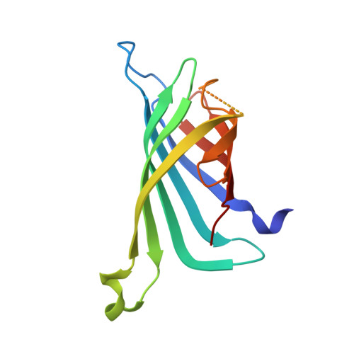
Structural consequences of cutting a binding loop: two circularly permuted variants of streptavidin.

Le Trong, I.Chu, V.Xing, Y.Lybrand, T.P.Stayton, P.S.Stenkamp, R.E.

(2013) Acta Crystallogr D Biol Crystallogr 69: 968-977

  • DOI: https://doi.org/10.1107/S0907444913003855
  • Primary Citation of Related Structures:  
    4GD9, 4GDA

  • PubMed Abstract: 

    Circular permutation of streptavidin was carried out in order to investigate the role of a main-chain amide in stabilizing the high-affinity complex of the protein and biotin. Mutant proteins CP49/48 and CP50/49 were constructed to place new N-termini at residues 49 and 50 in a flexible loop involved in stabilizing the biotin complex. Crystal structures of the two mutants show that half of each loop closes over the binding site, as observed in wild-type streptavidin, while the other half adopts the open conformation found in the unliganded state. The structures are consistent with kinetic and thermodynamic data and indicate that the loop plays a role in enthalpic stabilization of the bound state via the Asn49 amide-biotin hydrogen bond. In wild-type streptavidin, the entropic penalties of immobilizing a flexible portion of the protein to enhance binding are kept to a manageable level by using a contiguous loop of medium length (six residues) which is already constrained by its anchorage to strands of the β-barrel protein. A molecular-dynamics simulation for CP50/49 shows that cleavage of the binding loop results in increased structural fluctuations for Ser45 and that these fluctuations destabilize the streptavidin-biotin complex.


  • Organizational Affiliation

    Department of Biological Structure, University of Washington, Box 357420, Seattle, WA 98195-7420, USA.


Macromolecules
Find similar proteins by:  (by identity cutoff)  |  3D Structure
Entity ID: 1
MoleculeChains Sequence LengthOrganismDetailsImage
Streptavidin
A, B, C, D
132Streptomyces avidiniiMutation(s): 0 
UniProt
Find proteins for P22629 (Streptomyces avidinii)
Explore P22629 
Go to UniProtKB:  P22629
Entity Groups  
Sequence Clusters30% Identity50% Identity70% Identity90% Identity95% Identity100% Identity
UniProt GroupP22629
Sequence Annotations
Expand
  • Reference Sequence
Experimental Data & Validation

Experimental Data

  • Method: X-RAY DIFFRACTION
  • Resolution: 1.50 Å
  • R-Value Free: 0.230 
  • R-Value Work: 0.201 
  • R-Value Observed: 0.202 
  • Space Group: P 21 21 21
Unit Cell:
Length ( Å )Angle ( ˚ )
a = 54.72α = 90
b = 85.89β = 90
c = 100.14γ = 90
Software Package:
Software NamePurpose
DENZOdata reduction
SCALEPACKdata scaling
REFMACrefinement
PDB_EXTRACTdata extraction
Blu-Icedata collection
BALBESphasing

Structure Validation

View Full Validation Report


Ligand Structure Quality Assessment 


Entry History 

Deposition Data

Revision History  (Full details and data files)

  • Version 1.0: 2013-06-05
    Type: Initial release
  • Version 1.1: 2017-08-02
    Changes: Refinement description, Source and taxonomy
  • Version 1.2: 2017-11-15
    Changes: Refinement description, Structure summary
  • Version 1.3: 2023-09-13
    Changes: Data collection, Database references, Derived calculations, Refinement description