


Crystal Structure of the ERK2 complexed with E57
Kang, Y.N., Stuckey, J.A., Xie, X.To be published.
Experimental Data Snapshot
Entity ID: 1 | |||||
|---|---|---|---|---|---|
| Molecule | Chains | Sequence Length | Organism | Details | Image |
| Mitogen-activated protein kinase 1 | 360 | Homo sapiens | Mutation(s): 0 Gene Names: ERK2, MAPK1, PRKM1, PRKM2 EC: 2.7.11.24 | 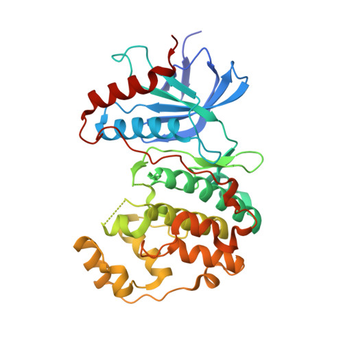 | |
UniProt & NIH Common Fund Data Resources | |||||
Find proteins for P28482 (Homo sapiens) Explore P28482 Go to UniProtKB: P28482 | |||||
PHAROS: P28482 GTEx: ENSG00000100030 | |||||
Entity Groups | |||||
| Sequence Clusters | 30% Identity50% Identity70% Identity90% Identity95% Identity100% Identity | ||||
| UniProt Group | P28482 | ||||
Sequence AnnotationsExpand | |||||
| |||||
| Ligands 3 Unique | |||||
|---|---|---|---|---|---|
| ID | Chains | Name / Formula / InChI Key | 2D Diagram | 3D Interactions | |
| E57 Query on E57 | B [auth A] | N-[(1S)-1-(3-chlorophenyl)-2-hydroxyethyl]-4-(2-{[(2S)-1-hydroxybutan-2-yl]amino}-5-methylpyrimidin-4-yl)-1H-pyrrole-2-carboxamide C22 H26 Cl N5 O3 GFHDSVUCETUKHU-PKOBYXMFSA-N |  | ||
| SO4 Query on SO4 | C [auth A], D [auth A], E [auth A] | SULFATE ION O4 S QAOWNCQODCNURD-UHFFFAOYSA-L |  | ||
| EDO Query on EDO | F [auth A] | 1,2-ETHANEDIOL C2 H6 O2 LYCAIKOWRPUZTN-UHFFFAOYSA-N |  | ||
| Length ( Å ) | Angle ( ˚ ) |
|---|---|
| a = 44.141 | α = 90 |
| b = 70.828 | β = 90 |
| c = 120.524 | γ = 90 |
| Software Name | Purpose |
|---|---|
| SCALEPACK | data scaling |
| BUSTER-TNT | refinement |
| PDB_EXTRACT | data extraction |
| BUSTER | refinement |
RCSB PDB (citation) is hosted by
RCSB PDB is a member of the

