4FBX

Complex structure of human protein kinase CK2 catalytic subunit crystallized in the presence of a bisubstrate inhibitor


Experimental Data Snapshot

  • Method: X-RAY DIFFRACTION
  • Resolution: 2.33 Å
  • R-Value Free: 0.249 
  • R-Value Work: 0.199 
  • R-Value Observed: 0.202 

wwPDB Validation   3D Report Full Report


This is version 1.3 of the entry. See complete history


Literature

A subnanomolar fluorescent probe for protein kinase CK2 interaction studies.

Enkvist, E.Viht, K.Bischoff, N.Vahter, J.Saaver, S.Raidaru, G.Issinger, O.G.Niefind, K.Uri, A.

(2012) Org Biomol Chem 10: 8645-8653

  • DOI: https://doi.org/10.1039/c2ob26022k
  • Primary Citation of Related Structures:  
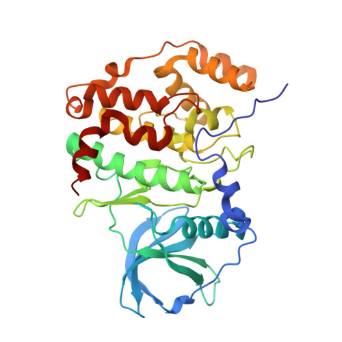
    4FBX

  • PubMed Abstract: 

    Up-regulation of an acidophilic protein kinase, CK2, has been established in several types of cancer. This cognition has made CK2 an important target for drug development for cancer chemotherapy. The characterization of potential drug candidates, determination of the structure and clarification of the functions of CK2 could be facilitated by the application of small-molecule fluorescent probes that bind to the active site of the enzyme with high affinity and selectivity. We have used a bisubstrate approach for the development of a highly potent inhibitor of CK2. 4,5,6,7-Tetrabromo-1H-benzimidazole was conjugated with peptides containing multiple aspartate residues via different linkers. The design of the inhibitors was by crystallographic analysis of the complex of an inhibitor with the catalytic subunit of the enzyme (CK2α). The inhibitory potency of the synthesized compounds was established in a kinetic assay that used thin layer chromatography for the measurement of the rate of phosphorylation of fluorescently labelled peptide 5-TAMRA-RADDSDDDDD. The most potent inhibitor, ARC-1502 (K(i) = 0.5 nM), revealed high selectivity for CK2α in a panel of 140 protein kinases. Labelling of ARC-1502 with PromoFluor-647 gave the fluorescent probe ARC-1504 that possessed subnanomolar affinity towards both CK2α and the holoenzyme. The probe was used in a fluorescence anisotropy-based binding assay to measure the concentration of CK2α and characterize non-labelled ligands binding to the active site of CK2α.


  • Organizational Affiliation

    Institute of Chemistry, University of Tartu, 14A Ravila St., 50411 Tartu, Estonia.


Macromolecules
Find similar proteins by:  (by identity cutoff)  |  3D Structure
Entity ID: 1
MoleculeChains Sequence LengthOrganismDetailsImage
Casein kinase II subunit alpha335Homo sapiensMutation(s): 0 
Gene Names: CSNK2A1CK2A1
EC: 2.7.11.1
UniProt & NIH Common Fund Data Resources
Find proteins for P68400 (Homo sapiens)
Explore P68400 
Go to UniProtKB:  P68400
PHAROS:  P68400
GTEx:  ENSG00000101266 
Entity Groups  
Sequence Clusters30% Identity50% Identity70% Identity90% Identity95% Identity100% Identity
UniProt GroupP68400
Sequence Annotations
Expand
  • Reference Sequence

Find similar proteins by:  Sequence   |   3D Structure  

Entity ID: 2
MoleculeChains Sequence LengthOrganismDetailsImage
bisubstrate inhibitor7N/AMutation(s): 0 
Sequence Annotations
Expand
  • Reference Sequence
Biologically Interesting Molecules (External Reference) 1 Unique
Experimental Data & Validation

Experimental Data

  • Method: X-RAY DIFFRACTION
  • Resolution: 2.33 Å
  • R-Value Free: 0.249 
  • R-Value Work: 0.199 
  • R-Value Observed: 0.202 
  • Space Group: P 43 21 2
Unit Cell:
Length ( Å )Angle ( ˚ )
a = 72.114α = 90
b = 72.114β = 90
c = 135.056γ = 90
Software Package:
Software NamePurpose
PHENIXmodel building
PHENIXrefinement
XDSdata reduction
SCALAdata scaling
PHENIXphasing

Structure Validation

View Full Validation Report


Entry History 

Deposition Data

Revision History  (Full details and data files)

  • Version 1.0: 2012-10-17
    Type: Initial release
  • Version 1.1: 2012-10-31
    Changes: Database references
  • Version 1.2: 2012-12-12
    Changes: Other
  • Version 1.3: 2024-02-28
    Changes: Data collection, Database references, Derived calculations