4F2K

Macrophage Migration Inhibitory Factor covalently complexed with phenethylisothiocyanate


Experimental Data Snapshot

  • Method: X-RAY DIFFRACTION
  • Resolution: 1.53 Å
  • R-Value Free: 0.228 
  • R-Value Work: 0.188 
  • R-Value Observed: 0.190 

wwPDB Validation   3D Report Full Report


Ligand Structure Quality Assessment 


This is version 1.2 of the entry. See complete history


Literature

Macrophage migration inhibitory factor covalently complexed with phenethyl isothiocyanate

Tyndall, J.D.A.Lue, H.Rutledge, M.T.Bernhagen, J.Hampton, M.B.Wilbanks, S.M.

(2012) Acta Crystallogr Sect F Struct Biol Cryst Commun 68: 999-1002

  • DOI: https://doi.org/10.1107/S1744309112030552
  • Primary Citation of Related Structures:  
    4F2K

  • PubMed Abstract: 

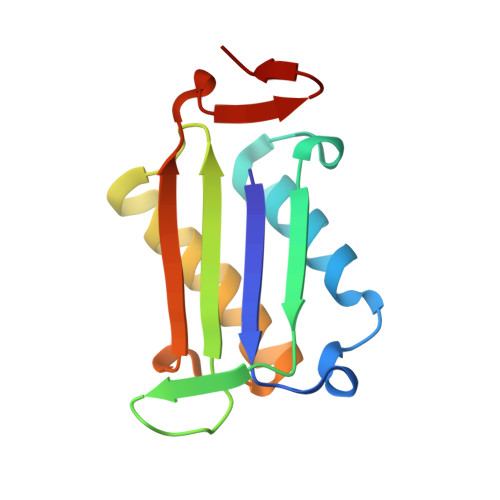
    Macrophage migration inhibitory factor is irreversibly inhibited via covalent modification by phenethyl isothiocyanate, a naturally occurring compound with anti-inflammatory and anticancer properties. The structure of the modified protein obtained from X-ray diffraction data to 1.64 Å resolution is presented. The inhibitor sits within a deep hydrophobic pocket between subunits of the homotrimer and is highly ordered. The secondary structure of macrophage migratory inhibitory factor is unchanged by this modification, but there are significant rearrangements, including of the side-chain position of Tyr37 and the main chain of residues 31-34. These changes may explain the decreased binding of the modified protein to the receptor CD74. Together with the pocket, the areas of conformational change define specific targets for the design of more selective and potent inhibitors as potential therapeutics.


  • Organizational Affiliation

    School of Pharmacy, University of Otago, PO Box 56, Dunedin 9054, New Zealand.


Macromolecules
Find similar proteins by:  (by identity cutoff)  |  3D Structure
Entity ID: 1
MoleculeChains Sequence LengthOrganismDetailsImage
Macrophage migration inhibitory factor
A, B, C
114Homo sapiensMutation(s): 0 
Gene Names: GLIFMIFMMIF
EC: 5.3.2.1 (PDB Primary Data), 5.3.3.12 (PDB Primary Data)
UniProt & NIH Common Fund Data Resources
Find proteins for P14174 (Homo sapiens)
Explore P14174 
Go to UniProtKB:  P14174
PHAROS:  P14174
GTEx:  ENSG00000240972 
Entity Groups  
Sequence Clusters30% Identity50% Identity70% Identity90% Identity95% Identity100% Identity
UniProt GroupP14174
Sequence Annotations
Expand
  • Reference Sequence
Small Molecules
Ligands 3 Unique
IDChains Name / Formula / InChI Key2D Diagram3D Interactions
LE2
Query on LE2

Download Ideal Coordinates CCD File 
D [auth A],
G [auth B],
K [auth C]
N-(2-phenylethyl)thioformamide
C9 H11 N S
WVJCPUDEWCGDGL-UHFFFAOYSA-N
SO4
Query on SO4

Download Ideal Coordinates CCD File 
E [auth A],
H [auth B],
L [auth C],
M [auth C]
SULFATE ION
O4 S
QAOWNCQODCNURD-UHFFFAOYSA-L
IPA
Query on IPA

Download Ideal Coordinates CCD File 
F [auth A],
I [auth B],
J [auth B],
N [auth C]
ISOPROPYL ALCOHOL
C3 H8 O
KFZMGEQAYNKOFK-UHFFFAOYSA-N
Experimental Data & Validation

Experimental Data

  • Method: X-RAY DIFFRACTION
  • Resolution: 1.53 Å
  • R-Value Free: 0.228 
  • R-Value Work: 0.188 
  • R-Value Observed: 0.190 
  • Space Group: P 21 21 21
Unit Cell:
Length ( Å )Angle ( ˚ )
a = 67.728α = 90
b = 68.245β = 90
c = 88.715γ = 90
Software Package:
Software NamePurpose
PHASERphasing
REFMACrefinement
PDB_EXTRACTdata extraction
CrystalCleardata collection
CrystalCleardata reduction
CrystalCleardata scaling

Structure Validation

View Full Validation Report


Ligand Structure Quality Assessment 


Entry History 

Deposition Data

Revision History  (Full details and data files)

  • Version 1.0: 2012-06-06
    Type: Initial release
  • Version 1.1: 2012-12-26
    Changes: Database references
  • Version 1.2: 2023-11-08
    Changes: Data collection, Database references, Derived calculations, Refinement description