


Crystal structure of a HLA-B associated transcript 3 (BAT3) from Homo sapiens at 1.80 A resolution
Joint Center for Structural Genomics (JCSG), Partnership for T-Cell Biology (TCELL)To be published.
Experimental Data Snapshot
wwPDB Validation 3D Report Full Report

Entity ID: 1 | |||||
|---|---|---|---|---|---|
| Molecule | Chains | Sequence Length | Organism | Details | Image |
| HLA-B-associated transcript 3 | 90 | Homo sapiens | Mutation(s): 0 Gene Names: BAG6, BAT3, BC003133, G3 | 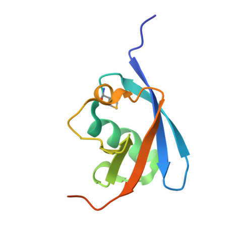 | |
UniProt & NIH Common Fund Data Resources | |||||
Find proteins for P46379 (Homo sapiens) Explore P46379 Go to UniProtKB: P46379 | |||||
PHAROS: P46379 GTEx: ENSG00000204463 | |||||
Entity Groups | |||||
| Sequence Clusters | 30% Identity50% Identity70% Identity90% Identity95% Identity100% Identity | ||||
| UniProt Group | P46379 | ||||
Sequence AnnotationsExpand | |||||
| |||||
| Ligands 1 Unique | |||||
|---|---|---|---|---|---|
| ID | Chains | Name / Formula / InChI Key | 2D Diagram | 3D Interactions | |
| SO4 Query on SO4 | C [auth A], D [auth A], E [auth B], F [auth B] | SULFATE ION O4 S QAOWNCQODCNURD-UHFFFAOYSA-L |  | ||
| Modified Residues 1 Unique | |||||
|---|---|---|---|---|---|
| ID | Chains | Type | Formula | 2D Diagram | Parent |
| MSE Query on MSE | A, B | L-PEPTIDE LINKING | C5 H11 N O2 Se |  | MET |
| Length ( Å ) | Angle ( ˚ ) |
|---|---|
| a = 61.32 | α = 90 |
| b = 74.09 | β = 90 |
| c = 43.119 | γ = 90 |
| Software Name | Purpose |
|---|---|
| MolProbity | model building |
| PDB_EXTRACT | data extraction |
| SHELX | phasing |
| SHARP | phasing |
| XSCALE | data scaling |
| REFMAC | refinement |
| XDS | data reduction |
| SHELXD | phasing |
RCSB PDB (citation) is hosted by
RCSB PDB is a member of the

