4DUU

The X-ray Crystal Structure of Full-Length type I Human Plasminogen


Experimental Data Snapshot

  • Method: X-RAY DIFFRACTION
  • Resolution: 5.20 Å
  • R-Value Free: 0.312 
  • R-Value Work: 0.268 
  • R-Value Observed: 0.270 

wwPDB Validation   3D Report Full Report


This is version 1.3 of the entry. See complete history


Literature

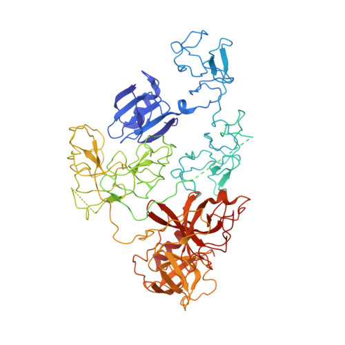
The X-ray crystal structure of full-length human plasminogen

Law, R.H.P.Caradoc-Davies, T.Cowieson, N.Horvath, A.J.Quek, A.J.Encarnacao, J.A.Steer, D.Cowan, A.Zhang, Q.Lu, B.G.C.Pike, R.N.Smith, A.I.Coughlin, P.B.Whisstock, J.C.

(2012) Cell Rep 1: 185-190

  • DOI: https://doi.org/10.1016/j.celrep.2012.02.012
  • Primary Citation of Related Structures:  
    4DUR, 4DUU

  • PubMed Abstract: 

    Plasminogen is the proenzyme precursor of the primary fibrinolytic protease plasmin. Circulating plasminogen, which comprises a Pan-apple (PAp) domain, five kringle domains (KR1-5), and a serine protease (SP) domain, adopts a closed, activation-resistant conformation. The kringle domains mediate interactions with fibrin clots and cell-surface receptors. These interactions trigger plasminogen to adopt an open form that can be cleaved and converted to plasmin by tissue-type and urokinase-type plasminogen activators. Here, the structure of closed plasminogen reveals that the PAp and SP domains, together with chloride ions, maintain the closed conformation through interactions with the kringle array. Differences in glycosylation alter the position of KR3, although in all structures the loop cleaved by plasminogen activators is inaccessible. The ligand-binding site of KR1 is exposed and likely governs proenzyme recruitment to targets. Furthermore, analysis of our structure suggests that KR5 peeling away from the PAp domain may initiate plasminogen conformational change.


  • Organizational Affiliation

    Department of Biochemistry and Molecular Biology, Monash University, Clayton, Melbourne, VIC 3800 Australia.


Macromolecules
Find similar proteins by:  (by identity cutoff)  |  3D Structure
Entity ID: 1
MoleculeChains Sequence LengthOrganismDetailsImage
Plasminogen791Homo sapiensMutation(s): 0 
EC: 3.4.21.7
UniProt & NIH Common Fund Data Resources
Find proteins for P00747 (Homo sapiens)
Explore P00747 
Go to UniProtKB:  P00747
PHAROS:  P00747
GTEx:  ENSG00000122194 
Entity Groups  
Sequence Clusters30% Identity50% Identity70% Identity90% Identity95% Identity100% Identity
UniProt GroupP00747
Sequence Annotations
Expand
  • Reference Sequence
Experimental Data & Validation

Experimental Data

  • Method: X-RAY DIFFRACTION
  • Resolution: 5.20 Å
  • R-Value Free: 0.312 
  • R-Value Work: 0.268 
  • R-Value Observed: 0.270 
  • Space Group: P 43 21 2
Unit Cell:
Length ( Å )Angle ( ˚ )
a = 110.12α = 90
b = 110.12β = 90
c = 234.815γ = 90
Software Package:
Software NamePurpose
ADSCdata collection
BUSTERrefinement
XDSdata reduction
SCALAdata scaling

Structure Validation

View Full Validation Report


Entry History 

Deposition Data

Revision History  (Full details and data files)

  • Version 1.0: 2012-03-28
    Type: Initial release
  • Version 1.1: 2012-04-04
    Changes: Structure summary
  • Version 1.2: 2013-06-26
    Changes: Database references
  • Version 1.3: 2023-11-08
    Changes: Data collection, Database references, Refinement description