4DRW

Crystal Structure of the Ternary Complex between S100A10, an Annexin A2 N-terminal Peptide and an AHNAK Peptide


Experimental Data Snapshot

  • Method: X-RAY DIFFRACTION
  • Resolution: 3.50 Å
  • R-Value Free: 0.327 
  • R-Value Work: 0.296 
  • R-Value Observed: 0.322 

wwPDB Validation   3D Report Full Report


This is version 1.3 of the entry. See complete history


Literature

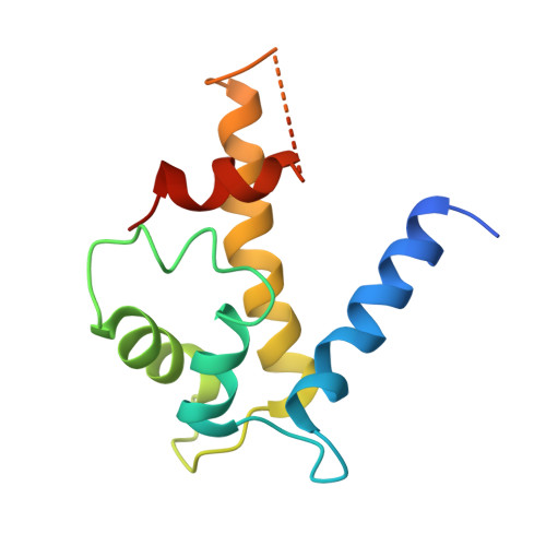
Structure of an asymmetric ternary protein complex provides insight for membrane interaction.

Dempsey, B.R.Rezvanpour, A.Lee, T.W.Barber, K.R.Junop, M.S.Shaw, G.S.

(2012) Structure 20: 1737-1745

  • DOI: https://doi.org/10.1016/j.str.2012.08.004
  • Primary Citation of Related Structures:  
    4DRW

  • PubMed Abstract: 

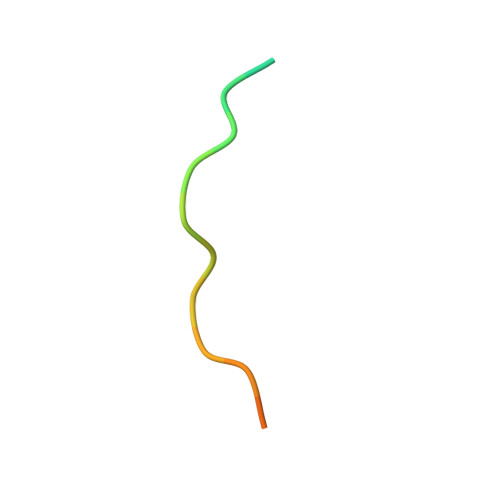
    Plasma membrane repair involves the coordinated effort of proteins and the inner phospholipid surface to mend the rupture and return the cell back to homeostasis. Here, we present the three-dimensional structure of a multiprotein complex that includes S100A10, annexin A2, and AHNAK, which along with dysferlin, functions in muscle and cardiac tissue repair. The 3.5 Å resolution X-ray structure shows that a single region from the AHNAK C terminus is recruited by an S100A10-annexin A2 heterotetramer, forming an asymmetric ternary complex. The AHNAK peptide adopts a coil conformation that arches across the heterotetramer contacting both annexin A2 and S100A10 protomers with tight affinity (∼30 nM) and establishing a structural rationale whereby both S100A10 and annexin proteins are needed in AHNAK recruitment. The structure evokes a model whereby AHNAK is targeted to the membrane surface through sandwiching of the binding region between the S100A10/annexin A2 complex and the phospholipid membrane.


  • Organizational Affiliation

    Department of Biochemistry, The University of Western Ontario, London, Ontario N6A 5C1, Canada.


Macromolecules
Find similar proteins by:  (by identity cutoff)  |  3D Structure
Entity ID: 1
MoleculeChains Sequence LengthOrganismDetailsImage
Protein S100-A10/Annexin A2 chimeric protein
A, B, C, D
121Homo sapiensMutation(s): 1 
Gene Names: S100A10ANX2LGCAL1LCLP11
UniProt & NIH Common Fund Data Resources
Find proteins for P60903 (Homo sapiens)
Explore P60903 
Go to UniProtKB:  P60903
PHAROS:  P60903
GTEx:  ENSG00000197747 
Find proteins for P07355 (Homo sapiens)
Explore P07355 
Go to UniProtKB:  P07355
PHAROS:  P07355
GTEx:  ENSG00000182718 
Entity Groups  
Sequence Clusters30% Identity50% Identity70% Identity90% Identity95% Identity100% Identity
UniProt GroupsP07355P60903
Sequence Annotations
Expand
  • Reference Sequence

Find similar proteins by:  Sequence   |   3D Structure  

Entity ID: 2
MoleculeChains Sequence LengthOrganismDetailsImage
Neuroblast differentiation-associated protein AHNAK
E, F
20Homo sapiensMutation(s): 0 
UniProt & NIH Common Fund Data Resources
Find proteins for Q09666 (Homo sapiens)
Explore Q09666 
Go to UniProtKB:  Q09666
PHAROS:  Q09666
GTEx:  ENSG00000124942 
Entity Groups  
Sequence Clusters30% Identity50% Identity70% Identity90% Identity95% Identity100% Identity
UniProt GroupQ09666
Sequence Annotations
Expand
  • Reference Sequence
Experimental Data & Validation

Experimental Data

  • Method: X-RAY DIFFRACTION
  • Resolution: 3.50 Å
  • R-Value Free: 0.327 
  • R-Value Work: 0.296 
  • R-Value Observed: 0.322 
  • Space Group: P 1
Unit Cell:
Length ( Å )Angle ( ˚ )
a = 47.755α = 71.62
b = 47.791β = 71.63
c = 62.817γ = 68.62
Software Package:
Software NamePurpose
CBASSdata collection
PHASERphasing
CNSrefinement
HKL-2000data reduction
HKL-2000data scaling

Structure Validation

View Full Validation Report


Entry History 

Deposition Data

Revision History  (Full details and data files)

  • Version 1.0: 2012-10-24
    Type: Initial release
  • Version 1.1: 2012-10-31
    Changes: Database references
  • Version 1.2: 2017-07-26
    Changes: Refinement description, Source and taxonomy
  • Version 1.3: 2023-09-13
    Changes: Data collection, Database references, Refinement description