4DNE

Crystal structure of a triple-mutant of streptavidin in complex with desthiobiotin


Experimental Data Snapshot

  • Method: X-RAY DIFFRACTION
  • Resolution: 1.88 Å
  • R-Value Free: 0.219 
  • R-Value Work: 0.181 
  • R-Value Observed: 0.183 

wwPDB Validation   3D Report Full Report


Ligand Structure Quality Assessment 


This is version 1.3 of the entry. See complete history


Literature

Contamination from an affinity column: an encounter with a new villain in the world of membrane-protein crystallization.

Panwar, P.Deniaud, A.Pebay-Peyroula, E.

(2012) Acta Crystallogr D Biol Crystallogr 68: 1272-1277

  • DOI: https://doi.org/10.1107/S090744491202639X
  • Primary Citation of Related Structures:  
    4DNE

  • PubMed Abstract: 

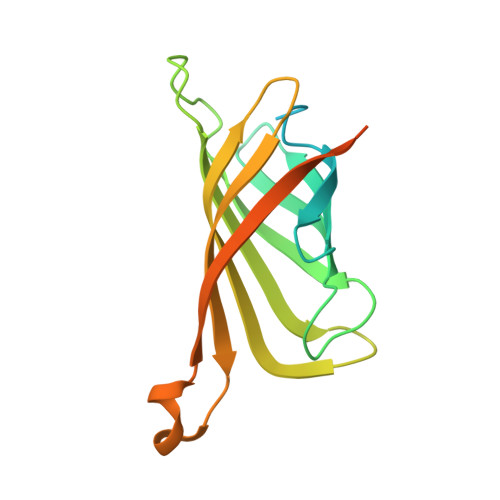
    Attempts to crystallize AtNTT1, a chloroplast ATP/ADP transporter from Arabidopsis thaliana, revealed an unexpected contaminant, Strep-Tactin, a variant of streptavidin that was used during purification of the protein. Although it was present in very small amounts, crystals of Strep-Tactin were reproducibly grown from the AtNTT1 solution. AtNTT1 was overexpressed in Escherichia coli and purified from detergent-solubilized membrane fractions using Strep-Tactin affinity chromatography based on an engineered streptavidin. The contamination of protein solutions purified on Strep-Tactin columns has never been described previously and seems to be specific to membrane proteins solubilized in detergents. Trace amounts of Strep-Tactin were observed to be eluted from a Strep-Tactin column using several routinely used detergents, illustrating their possible role in the contamination. This finding raises an alarm and suggests caution in membrane-protein purification using Strep-Tactin affinity columns, where detergents are essential components. The small crystals of contaminant protein led to the structure at 1.9 Å resolution of Strep-Tactin in complex with desthiobiotin.


  • Organizational Affiliation

    CEA, Institut de Biologie Structurale Jean-Pierre Ebel, 41 Rue Jules Horowitz, 38027 Grenoble, France.


Macromolecules
Find similar proteins by:  (by identity cutoff)  |  3D Structure
Entity ID: 1
MoleculeChains Sequence LengthOrganismDetailsImage
Streptavidin
A, B
183Streptomyces avidiniiMutation(s): 3 
UniProt
Find proteins for P22629 (Streptomyces avidinii)
Explore P22629 
Go to UniProtKB:  P22629
Entity Groups  
Sequence Clusters30% Identity50% Identity70% Identity90% Identity95% Identity100% Identity
UniProt GroupP22629
Sequence Annotations
Expand
  • Reference Sequence
Experimental Data & Validation

Experimental Data

  • Method: X-RAY DIFFRACTION
  • Resolution: 1.88 Å
  • R-Value Free: 0.219 
  • R-Value Work: 0.181 
  • R-Value Observed: 0.183 
  • Space Group: I 2 2 2
Unit Cell:
Length ( Å )Angle ( ˚ )
a = 46.72α = 90
b = 93.782β = 90
c = 104.413γ = 90
Software Package:
Software NamePurpose
MxCuBEdata collection
MERLOTphasing
REFMACrefinement
XDSdata reduction
SCALAdata scaling

Structure Validation

View Full Validation Report


Ligand Structure Quality Assessment 


Entry History 

Deposition Data

Revision History  (Full details and data files)

  • Version 1.0: 2012-09-26
    Type: Initial release
  • Version 1.1: 2013-01-23
    Changes: Database references
  • Version 1.2: 2016-04-06
    Changes: Non-polymer description
  • Version 1.3: 2023-09-13
    Changes: Data collection, Database references, Derived calculations, Refinement description