4DK7

Crystal structure of LXR ligand binding domain in complex with full agonist 1


Experimental Data Snapshot

  • Method: X-RAY DIFFRACTION
  • Resolution: 2.45 Å
  • R-Value Free: 0.270 
  • R-Value Work: 0.230 
  • R-Value Observed: 0.276 

wwPDB Validation   3D Report Full Report


Ligand Structure Quality Assessment 


This is version 1.1 of the entry. See complete history


Literature

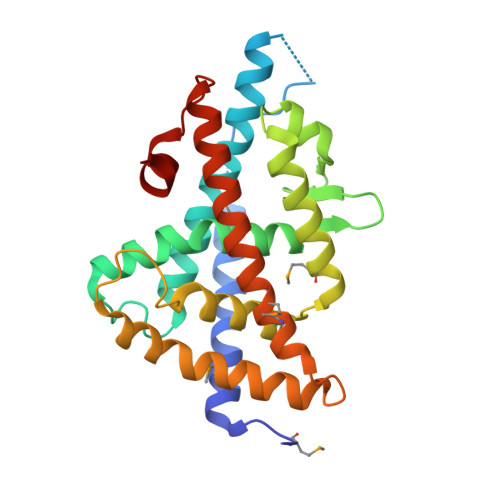
Discovery of a new binding mode for a series of liver X receptor agonists.

Kopecky, D.J.Jiao, X.Y.Fisher, B.McKendry, S.Labelle, M.Piper, D.E.Coward, P.Shiau, A.K.Escaron, P.Danao, J.Chai, A.Jaen, J.Kayser, F.

(2012) Bioorg Med Chem Lett 22: 2407-2410

  • DOI: https://doi.org/10.1016/j.bmcl.2012.02.028
  • Primary Citation of Related Structures:  
    4DK7, 4DK8

  • PubMed Abstract: 

    Structural modification of a series of dual LXRα/β agonists led to the identification of a new class of LXRβ partial agonists. An X-ray co-crystal structure shows that a representative member of this series, pyrrole 5, binds to LXRβ with a reversed orientation compared to 1.


  • Organizational Affiliation

    Department of Medicinal Chemistry, Amgen Inc., 1120 Veterans Blvd., South San Francisco, CA 94080, USA. dkopecky@amgen.com


Macromolecules
Find similar proteins by:  (by identity cutoff)  |  3D Structure
Entity ID: 1
MoleculeChains Sequence LengthOrganismDetailsImage
Oxysterols receptor LXR-beta
A, C
247Homo sapiensMutation(s): 0 
Gene Names: NR1H2LXRBNERUNR
UniProt & NIH Common Fund Data Resources
Find proteins for P55055 (Homo sapiens)
Explore P55055 
Go to UniProtKB:  P55055
PHAROS:  P55055
GTEx:  ENSG00000131408 
Entity Groups  
Sequence Clusters30% Identity50% Identity70% Identity90% Identity95% Identity100% Identity
UniProt GroupP55055
Sequence Annotations
Expand
  • Reference Sequence

Find similar proteins by:  Sequence   |   3D Structure  

Entity ID: 2
MoleculeChains Sequence LengthOrganismDetailsImage
Nuclear receptor coactivator 1
B, D
12Homo sapiensMutation(s): 0 
EC: 2.3.1.48
UniProt & NIH Common Fund Data Resources
Find proteins for Q15788 (Homo sapiens)
Explore Q15788 
Go to UniProtKB:  Q15788
PHAROS:  Q15788
GTEx:  ENSG00000084676 
Entity Groups  
Sequence Clusters30% Identity50% Identity70% Identity90% Identity95% Identity100% Identity
UniProt GroupQ15788
Sequence Annotations
Expand
  • Reference Sequence
Small Molecules
Ligands 3 Unique
IDChains Name / Formula / InChI Key2D Diagram3D Interactions
0KS
Query on 0KS

Download Ideal Coordinates CCD File 
E [auth A],
L [auth C]
N-[4-(1,1,1,3,3,3-hexafluoro-2-hydroxypropan-2-yl)phenyl]-N-methylbenzenesulfonamide
C16 H13 F6 N O3 S
CNVKZYLQZYULJV-UHFFFAOYSA-N
ACT
Query on ACT

Download Ideal Coordinates CCD File 
F [auth A]ACETATE ION
C2 H3 O2
QTBSBXVTEAMEQO-UHFFFAOYSA-M
CA
Query on CA

Download Ideal Coordinates CCD File 
G [auth A]
H [auth A]
I [auth A]
J [auth A]
K [auth A]
G [auth A],
H [auth A],
I [auth A],
J [auth A],
K [auth A],
M [auth C],
N [auth C]
CALCIUM ION
Ca
BHPQYMZQTOCNFJ-UHFFFAOYSA-N
Modified Residues  1 Unique
IDChains TypeFormula2D DiagramParent
MSE
Query on MSE
A, C
L-PEPTIDE LINKINGC5 H11 N O2 SeMET
Binding Affinity Annotations 
IDSourceBinding Affinity
0KS PDBBind:  4DK7 IC50: 20 (nM) from 1 assay(s)
Binding MOAD:  4DK7 IC50: 20 (nM) from 1 assay(s)
BindingDB:  4DK7 IC50: 20 (nM) from 1 assay(s)
Experimental Data & Validation

Experimental Data

  • Method: X-RAY DIFFRACTION
  • Resolution: 2.45 Å
  • R-Value Free: 0.270 
  • R-Value Work: 0.230 
  • R-Value Observed: 0.276 
  • Space Group: P 43 21 2
Unit Cell:
Length ( Å )Angle ( ˚ )
a = 87.261α = 90
b = 87.261β = 90
c = 197.709γ = 90
Software Package:
Software NamePurpose
DENZOdata reduction
SCALEPACKdata scaling
CNSrefinement
PDB_EXTRACTdata extraction
CNSphasing

Structure Validation

View Full Validation Report


Ligand Structure Quality Assessment 


Entry History 

Deposition Data

Revision History  (Full details and data files)

  • Version 1.0: 2012-03-21
    Type: Initial release
  • Version 1.1: 2012-05-09
    Changes: Database references