Dynamics of the Peripheral Membrane Protein P2 from Human Myelin Measured by Neutron Scattering-A Comparison between Wild-Type Protein and a Hinge Mutant.
Laulumaa, S., Nieminen, T., Lehtimaki, M., Aggarwal, S., Simons, M., Koza, M.M., Vattulainen, I., Kursula, P., Natali, F.(2015) PLoS One 10: 28954
- PubMed: 26068118
- DOI: https://doi.org/10.1371/journal.pone.0128954
- Primary Citation of Related Structures:
4D6A, 4D6B - PubMed Abstract:
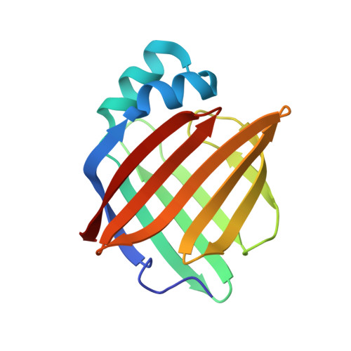
Myelin protein P2 is a fatty acid-binding structural component of the myelin sheath in the peripheral nervous system, and its function is related to its membrane binding capacity. Here, the link between P2 protein dynamics and structure and function was studied using elastic incoherent neutron scattering (EINS). The P38G mutation, at the hinge between the β barrel and the α-helical lid, increased the lipid stacking capacity of human P2 in vitro, and the mutated protein was also functional in cultured cells. The P38G mutation did not change the overall structure of the protein. For a deeper insight into P2 structure-function relationships, information on protein dynamics in the 10 ps to 1 ns time scale was obtained using EINS. Values of mean square displacements mainly from protein H atoms were extracted for wild-type P2 and the P38G mutant and compared. Our results show that at physiological temperatures, the P38G mutant is more dynamic than the wild-type P2 protein, especially on a slow 1-ns time scale. Molecular dynamics simulations confirmed the enhanced dynamics of the mutant variant, especially within the portal region in the presence of bound fatty acid. The increased softness of the hinge mutant of human myelin P2 protein is likely related to an enhanced flexibility of the portal region of this fatty acid-binding protein, as well as to its interactions with the lipid bilayer surface requiring conformational adaptations.
Organizational Affiliation:
Biochemistry and Molecular Medicine & Biocenter Oulu, University of Oulu, Oulu, Finland; German Electron Synchrotron (DESY), Hamburg, Germany; European Spallation Source (ESS), Lund, Sweden.