4CTH

Neprilysin variant G399V,G714K in complex with phosphoramidon


Experimental Data Snapshot

  • Method: X-RAY DIFFRACTION
  • Resolution: 2.15 Å
  • R-Value Free: 0.261 
  • R-Value Work: 0.209 
  • R-Value Observed: 0.212 

wwPDB Validation   3D Report Full Report


Ligand Structure Quality Assessment 


This is version 1.4 of the entry. See complete history


Literature

Engineering Neprilysin Activity and Specificity to Create a Novel Therapeutic for Alzheimer'S Disease.

Webster, C.I.Burrell, M.Olsson, L.Fowler, S.B.Digby, S.Sandercock, A.Snijder, A.Tebbe, J.Haupts, U.Grudzinska, J.Jermutus, L.Andersson, C.

(2014) PLoS One 9: 04001

  • DOI: https://doi.org/10.1371/journal.pone.0104001
  • Primary Citation of Related Structures:  
    4CTH

  • PubMed Abstract: 

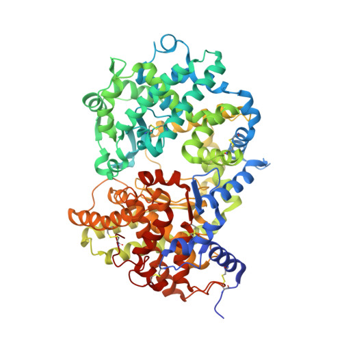
    Neprilysin is a transmembrane zinc metallopeptidase that degrades a wide range of peptide substrates. It has received attention as a potential therapy for Alzheimer's disease due to its ability to degrade the peptide amyloid beta. However, its broad range of peptide substrates has the potential to limit its therapeutic use due to degradation of additional peptides substrates that tightly regulate many physiological processes. We sought to generate a soluble version of the ectodomain of neprilysin with improved activity and specificity towards amyloid beta as a potential therapeutic for Alzheimer's disease. Extensive amino acid substitutions were performed at positions surrounding the active site and inner surface of the enzyme and variants screened for activity on amyloid beta 1-40, 1-42 and a variety of other physiologically relevant peptides. We identified several mutations that modulated and improved both enzyme selectivity and intrinsic activity. Neprilysin variant G399V/G714K displayed an approximately 20-fold improved activity on amyloid beta 1-40 and up to a 3,200-fold reduction in activity on other peptides. Along with the altered peptide substrate specificity, the mutant enzyme produced a markedly altered series of amyloid beta cleavage products compared to the wild-type enzyme. Crystallisation of the mutant enzyme revealed that the amino acid substitutions result in alteration of the shape and size of the pocket containing the active site compared to the wild-type enzyme. The mutant enzyme offers the potential for the more efficient degradation of amyloid beta in vivo as a therapeutic for the treatment of Alzheimer's disease.


  • Organizational Affiliation

    Antibody Discovery and Protein Engineering, MedImmune, Cambridge, United Kingdom.


Macromolecules
Find similar proteins by:  (by identity cutoff)  |  3D Structure
Entity ID: 1
MoleculeChains Sequence LengthOrganismDetailsImage
NEPRILYSIN709Homo sapiensMutation(s): 2 
EC: 3.4.24.11
UniProt & NIH Common Fund Data Resources
Find proteins for P08473 (Homo sapiens)
Explore P08473 
Go to UniProtKB:  P08473
PHAROS:  P08473
GTEx:  ENSG00000196549 
Entity Groups  
Sequence Clusters30% Identity50% Identity70% Identity90% Identity95% Identity100% Identity
UniProt GroupP08473
Sequence Annotations
Expand
  • Reference Sequence
Small Molecules
Ligands 4 Unique
IDChains Name / Formula / InChI Key2D Diagram3D Interactions
RDF
Query on RDF

Download Ideal Coordinates CCD File 
N [auth A]N-ALPHA-L-RHAMNOPYRANOSYLOXY(HYDROXYPHOSPHINYL)-L-LEUCYL-L-TRYPTOPHAN
C23 H34 N3 O10 P
ZPHBZEQOLSRPAK-XLCYBJAPSA-N
NAG
Query on NAG

Download Ideal Coordinates CCD File 
B [auth A],
C [auth A],
D [auth A]
2-acetamido-2-deoxy-beta-D-glucopyranose
C8 H15 N O6
OVRNDRQMDRJTHS-FMDGEEDCSA-N
GOL
Query on GOL

Download Ideal Coordinates CCD File 
F [auth A]
G [auth A]
H [auth A]
I [auth A]
J [auth A]
F [auth A],
G [auth A],
H [auth A],
I [auth A],
J [auth A],
K [auth A],
L [auth A],
M [auth A]
GLYCEROL
C3 H8 O3
PEDCQBHIVMGVHV-UHFFFAOYSA-N
ZN
Query on ZN

Download Ideal Coordinates CCD File 
E [auth A]ZINC ION
Zn
PTFCDOFLOPIGGS-UHFFFAOYSA-N
Binding Affinity Annotations 
IDSourceBinding Affinity
RDF BindingDB:  4CTH Ki: 4 (nM) from 1 assay(s)
IC50: min: 0.4, max: 200 (nM) from 13 assay(s)
Biologically Interesting Molecules (External Reference) 1 Unique
Experimental Data & Validation

Experimental Data

  • Method: X-RAY DIFFRACTION
  • Resolution: 2.15 Å
  • R-Value Free: 0.261 
  • R-Value Work: 0.209 
  • R-Value Observed: 0.212 
  • Space Group: P 32 2 1
Unit Cell:
Length ( Å )Angle ( ˚ )
a = 108.799α = 90
b = 108.799β = 90
c = 112.942γ = 120
Software Package:
Software NamePurpose
BUSTERrefinement
MOSFLMdata reduction
SCALAdata scaling
MOLREPphasing

Structure Validation

View Full Validation Report


Ligand Structure Quality Assessment 


Entry History 

Revision History  (Full details and data files)

  • Version 1.0: 2014-08-13
    Type: Initial release
  • Version 1.1: 2018-06-20
    Changes: Data collection, Structure summary
  • Version 1.2: 2019-05-08
    Changes: Data collection, Derived calculations, Experimental preparation
  • Version 1.3: 2020-07-29
    Type: Remediation
    Reason: Carbohydrate remediation
    Changes: Data collection, Derived calculations, Other, Structure summary
  • Version 1.4: 2023-12-20
    Changes: Data collection, Database references, Refinement description, Structure summary