4CFH

Structure of an active form of mammalian AMPK


Experimental Data Snapshot

  • Method: X-RAY DIFFRACTION
  • Resolution: 3.24 Å
  • R-Value Free: 0.268 
  • R-Value Work: 0.233 
  • R-Value Observed: 0.234 

wwPDB Validation   3D Report Full Report


This is version 1.3 of the entry. See complete history


Literature

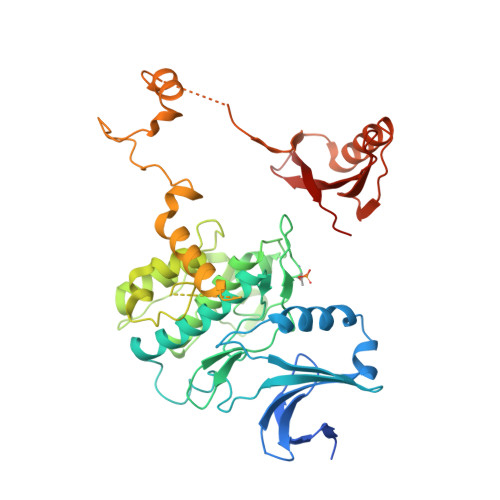
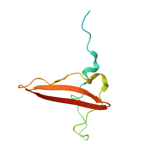
Structure of Mammalian Ampk and its Regulation by Adp

Xiao, B.Sanders, M.J.Underwood, E.Heath, R.Mayer, F.Carmena, D.Jing, C.Walker, P.A.Eccleston, J.F.Haire, L.F.Saiu, P.Howell, S.A.Aasland, R.Martin, S.R.Carling, D.Gamblin, S.J.

(2011) Nature 472: 230

  • DOI: https://doi.org/10.1038/nature09932
  • Primary Citation of Related Structures:  
    2Y8L, 2Y8Q, 2YA3, 4CFH

  • PubMed Abstract: 

    The heterotrimeric AMP-activated protein kinase (AMPK) has a key role in regulating cellular energy metabolism; in response to a fall in intracellular ATP levels it activates energy-producing pathways and inhibits energy-consuming processes. AMPK has been implicated in a number of diseases related to energy metabolism including type 2 diabetes, obesity and, most recently, cancer. AMPK is converted from an inactive form to a catalytically competent form by phosphorylation of the activation loop within the kinase domain: AMP binding to the γ-regulatory domain promotes phosphorylation by the upstream kinase, protects the enzyme against dephosphorylation, as well as causing allosteric activation. Here we show that ADP binding to just one of the two exchangeable AXP (AMP/ADP/ATP) binding sites on the regulatory domain protects the enzyme from dephosphorylation, although it does not lead to allosteric activation. Our studies show that active mammalian AMPK displays significantly tighter binding to ADP than to Mg-ATP, explaining how the enzyme is regulated under physiological conditions where the concentration of Mg-ATP is higher than that of ADP and much higher than that of AMP. We have determined the crystal structure of an active AMPK complex. The structure shows how the activation loop of the kinase domain is stabilized by the regulatory domain and how the kinase linker region interacts with the regulatory nucleotide-binding site that mediates protection against dephosphorylation. From our biochemical and structural data we develop a model for how the energy status of a cell regulates AMPK activity.


  • Organizational Affiliation

    MRC National Institute for Medical Research, The Ridgeway, Mill Hill, London NW7 1AA, UK.


Macromolecules
Find similar proteins by:  (by identity cutoff)  |  3D Structure
Entity ID: 1
MoleculeChains Sequence LengthOrganismDetailsImage
5'-AMP-ACTIVATED PROTEIN KINASE CATALYTIC SUBUNIT ALPHA-1493Rattus norvegicusMutation(s): 0 
EC: 2.7.11.1
UniProt
Find proteins for P54645 (Rattus norvegicus)
Explore P54645 
Go to UniProtKB:  P54645
Entity Groups  
Sequence Clusters30% Identity50% Identity70% Identity90% Identity95% Identity100% Identity
UniProt GroupP54645
Sequence Annotations
Expand
  • Reference Sequence
Find similar proteins by:  (by identity cutoff)  |  3D Structure
Entity ID: 2
MoleculeChains Sequence LengthOrganismDetailsImage
5'-AMP-ACTIVATED PROTEIN KINASE SUBUNIT BETA-287Homo sapiensMutation(s): 0 
UniProt & NIH Common Fund Data Resources
Find proteins for O43741 (Homo sapiens)
Explore O43741 
Go to UniProtKB:  O43741
PHAROS:  O43741
GTEx:  ENSG00000131791 
Entity Groups  
Sequence Clusters30% Identity50% Identity70% Identity90% Identity95% Identity100% Identity
UniProt GroupO43741
Sequence Annotations
Expand
  • Reference Sequence
Find similar proteins by:  (by identity cutoff)  |  3D Structure
Entity ID: 3
MoleculeChains Sequence LengthOrganismDetailsImage
5'-AMP-ACTIVATED PROTEIN KINASE CATALYTIC SUBUNIT ALPHA-127Rattus norvegicusMutation(s): 0 
EC: 2.7.11.1
UniProt
Find proteins for P54645 (Rattus norvegicus)
Explore P54645 
Go to UniProtKB:  P54645
Entity Groups  
Sequence Clusters30% Identity50% Identity70% Identity90% Identity95% Identity100% Identity
UniProt GroupP54645
Sequence Annotations
Expand
  • Reference Sequence
Find similar proteins by:  (by identity cutoff)  |  3D Structure
Entity ID: 4
MoleculeChains Sequence LengthOrganismDetailsImage
5'-AMP-ACTIVATED PROTEIN KINASE SUBUNIT GAMMA-1D [auth E]330Rattus norvegicusMutation(s): 0 
UniProt
Find proteins for P80385 (Rattus norvegicus)
Explore P80385 
Go to UniProtKB:  P80385
Entity Groups  
Sequence Clusters30% Identity50% Identity70% Identity90% Identity95% Identity100% Identity
UniProt GroupP80385
Sequence Annotations
Expand
  • Reference Sequence
Small Molecules
Modified Residues  1 Unique
IDChains TypeFormula2D DiagramParent
TPO
Query on TPO
A
L-PEPTIDE LINKINGC4 H10 N O6 PTHR
Experimental Data & Validation

Experimental Data

  • Method: X-RAY DIFFRACTION
  • Resolution: 3.24 Å
  • R-Value Free: 0.268 
  • R-Value Work: 0.233 
  • R-Value Observed: 0.234 
  • Space Group: P 41 21 2
Unit Cell:
Length ( Å )Angle ( ˚ )
a = 133.917α = 90
b = 133.917β = 90
c = 141.896γ = 90
Software Package:
Software NamePurpose
DENZOdata reduction
SCALEPACKdata scaling
AMoREphasing
PHASERphasing
PHENIXrefinement

Structure Validation

View Full Validation Report


Entry History 

Deposition Data

Revision History  (Full details and data files)

  • Version 1.0: 2013-12-25
    Type: Initial release
  • Version 1.1: 2014-01-08
    Changes: Database references
  • Version 1.2: 2019-05-08
    Changes: Data collection, Derived calculations, Experimental preparation, Other
  • Version 1.3: 2023-12-20
    Changes: Data collection, Database references, Derived calculations, Other, Refinement description