4BIK

Structure of a disulfide locked mutant of Intermedilysin with human CD59


Experimental Data Snapshot

  • Method: X-RAY DIFFRACTION
  • Resolution: 3.49 Å
  • R-Value Free: 0.266 
  • R-Value Work: 0.212 
  • R-Value Observed: 0.203 

wwPDB Validation   3D Report Full Report


This is version 1.3 of the entry. See complete history


Literature

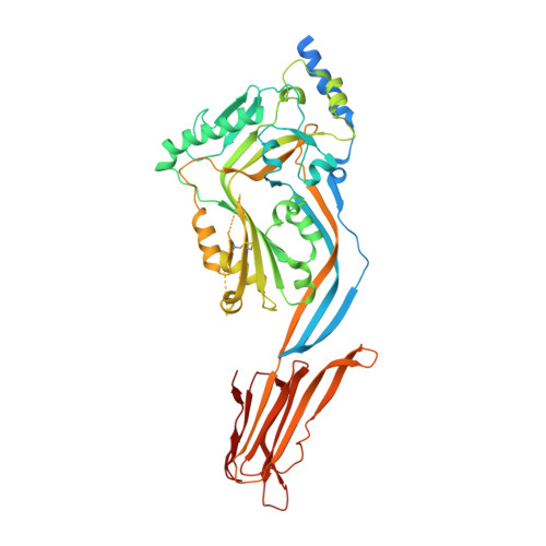
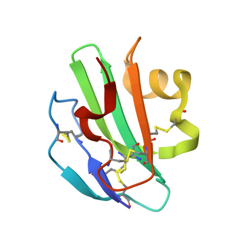
Structural Basis for Recognition of the Pore- Forming Toxin Intermedilysin by Human Complement Receptor Cd59

Johnson, S.Brooks, N.J.Smith, R.A.G.Lea, S.M.Bubeck, D.

(2013) Cell Rep 3: 1369

  • DOI: https://doi.org/10.1016/j.celrep.2013.04.029
  • Primary Citation of Related Structures:  
    4BIK

  • PubMed Abstract: 

    Pore-forming proteins containing the structurally conserved membrane attack complex/perforin fold play an important role in immunity and host-pathogen interactions. Intermedilysin (ILY) is an archetypal member of a cholesterol-dependent cytolysin subclass that hijacks the complement receptor CD59 to make cytotoxic pores in human cells. ILY directly competes for the membrane attack complex binding site on CD59, rendering cells susceptible to complement lysis. To understand how these bacterial pores form in lipid bilayers and the role CD59 plays in complement regulation, we determined the crystal structure of human CD59 bound to ILY. Here, we show the ILY-CD59 complex at 3.5 Å resolution and identify two interfaces mediating this host-pathogen interaction. An ILY-derived peptide based on the binding site inhibits pore formation in a CD59-containing liposome model system. These data provide insight into how CD59 coordinates ILY monomers, nucleating an early prepore state, and suggest a potential mechanism of inhibition for the complement terminal pathway.


  • Organizational Affiliation

    Sir William Dunn School of Pathology, University of Oxford, South Parks Road, Oxford OX1 3RE, UK.


Macromolecules
Find similar proteins by:  (by identity cutoff)  |  3D Structure
Entity ID: 1
MoleculeChains Sequence LengthOrganismDetailsImage
INTERMEDILYSIN
A, C
535Streptococcus intermediusMutation(s): 2 
Membrane Entity: Yes 
UniProt
Find proteins for Q9LCB8 (Streptococcus intermedius)
Explore Q9LCB8 
Go to UniProtKB:  Q9LCB8
Entity Groups  
Sequence Clusters30% Identity50% Identity70% Identity90% Identity95% Identity100% Identity
UniProt GroupQ9LCB8
Sequence Annotations
Expand
  • Reference Sequence
Find similar proteins by:  (by identity cutoff)  |  3D Structure
Entity ID: 2
MoleculeChains Sequence LengthOrganismDetailsImage
CD59 GLYCOPROTEIN
B, D
79Homo sapiensMutation(s): 0 
Membrane Entity: Yes 
UniProt & NIH Common Fund Data Resources
Find proteins for P13987 (Homo sapiens)
Explore P13987 
Go to UniProtKB:  P13987
PHAROS:  P13987
GTEx:  ENSG00000085063 
Entity Groups  
Sequence Clusters30% Identity50% Identity70% Identity90% Identity95% Identity100% Identity
UniProt GroupP13987
Sequence Annotations
Expand
  • Reference Sequence
Experimental Data & Validation

Experimental Data

  • Method: X-RAY DIFFRACTION
  • Resolution: 3.49 Å
  • R-Value Free: 0.266 
  • R-Value Work: 0.212 
  • R-Value Observed: 0.203 
  • Space Group: P 1
Unit Cell:
Length ( Å )Angle ( ˚ )
a = 27.4α = 63.75
b = 101.45β = 89.95
c = 112.88γ = 90.02
Software Package:
Software NamePurpose
PHENIXrefinement
XDSdata reduction
Aimlessdata scaling
PHASERphasing

Structure Validation

View Full Validation Report


Entry History 

Deposition Data

Revision History  (Full details and data files)

  • Version 1.0: 2013-05-08
    Type: Initial release
  • Version 1.1: 2013-05-29
    Changes: Database references
  • Version 1.2: 2013-06-19
    Changes: Database references
  • Version 1.3: 2023-12-20
    Changes: Data collection, Database references, Other, Refinement description