4AY9

Structure of follicle-stimulating hormone in complex with the entire ectodomain of its receptor


Experimental Data Snapshot

  • Method: X-RAY DIFFRACTION
  • Resolution: 2.50 Å
  • R-Value Free: 0.269 
  • R-Value Work: 0.235 
  • R-Value Observed: 0.237 

wwPDB Validation   3D Report Full Report


Ligand Structure Quality Assessment 


This is version 1.3 of the entry. See complete history


Literature

Structure of Follicle-Stimulating Hormone in Complex with the Entire Ectodomain of its Receptor.

Jiang, X.Liu, H.Chen, X.Chen, P.Fischer, D.Sriraman, V.Yu, H.N.Arkinstall, S.He, X.

(2012) Proc Natl Acad Sci U S A 109: 12491

  • DOI: https://doi.org/10.1073/pnas.1206643109
  • Primary Citation of Related Structures:  
    4AY9

  • PubMed Abstract: 

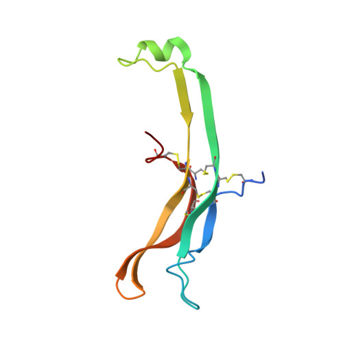
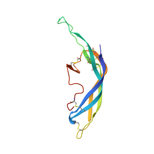
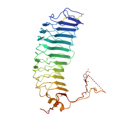
    FSH, a glycoprotein hormone, and the FSH receptor (FSHR), a G protein-coupled receptor, play central roles in human reproduction. We report the crystal structure of FSH in complex with the entire extracellular domain of FSHR (FSHR(ED)), including the enigmatic hinge region that is responsible for signal specificity. Surprisingly, the hinge region does not form a separate structural unit as widely anticipated but is part of the integral structure of FSHR(ED). In addition to the known hormone-binding site, FSHR(ED) provides interaction sites with the hormone: a sulfotyrosine (sTyr) site in the hinge region consistent with previous studies and a potential exosite resulting from putative receptor trimerization. Our structure, in comparison to others, suggests FSHR interacts with its ligand in two steps: ligand recruitment followed by sTyr recognition. FSH first binds to the high-affinity hormone-binding subdomain of FSHR and reshapes the ligand conformation to form a sTyr-binding pocket. FSHR then inserts its sTyr (i.e., sulfated Tyr335) into the FSH nascent pocket, eventually leading to receptor activation.


  • Organizational Affiliation

    EMD Serono Research Institute, Billerica, MA 01821, USA.


Macromolecules
Find similar proteins by:  (by identity cutoff)  |  3D Structure
Entity ID: 1
MoleculeChains Sequence LengthOrganismDetailsImage
GLYCOPROTEIN HORMONES, ALPHA POLYPEPTIDEA,
C [auth D],
E [auth G]
92Homo sapiensMutation(s): 0 
UniProt & NIH Common Fund Data Resources
Find proteins for P01215 (Homo sapiens)
Explore P01215 
Go to UniProtKB:  P01215
PHAROS:  P01215
GTEx:  ENSG00000135346 
Entity Groups  
Sequence Clusters30% Identity50% Identity70% Identity90% Identity95% Identity100% Identity
UniProt GroupP01215
Sequence Annotations
Expand
  • Reference Sequence
Find similar proteins by:  (by identity cutoff)  |  3D Structure
Entity ID: 2
MoleculeChains Sequence LengthOrganismDetailsImage
FOLLITROPIN SUBUNIT BETAB,
D [auth E],
F [auth H]
111Homo sapiensMutation(s): 0 
UniProt & NIH Common Fund Data Resources
Find proteins for P01225 (Homo sapiens)
Explore P01225 
Go to UniProtKB:  P01225
PHAROS:  P01225
GTEx:  ENSG00000131808 
Entity Groups  
Sequence Clusters30% Identity50% Identity70% Identity90% Identity95% Identity100% Identity
UniProt GroupP01225
Sequence Annotations
Expand
  • Reference Sequence
Find similar proteins by:  (by identity cutoff)  |  3D Structure
Entity ID: 3
MoleculeChains Sequence LengthOrganismDetailsImage
FOLLICLE-STIMULATING HORMONE RECEPTORG [auth X],
H [auth Y],
I [auth Z]
350Homo sapiensMutation(s): 0 
UniProt & NIH Common Fund Data Resources
Find proteins for P23945 (Homo sapiens)
Explore P23945 
Go to UniProtKB:  P23945
PHAROS:  P23945
GTEx:  ENSG00000170820 
Entity Groups  
Sequence Clusters30% Identity50% Identity70% Identity90% Identity95% Identity100% Identity
UniProt GroupP23945
Sequence Annotations
Expand
  • Reference Sequence
Small Molecules
Ligands 1 Unique
IDChains Name / Formula / InChI Key2D Diagram3D Interactions
NAG
Query on NAG

Download Ideal Coordinates CCD File 
J [auth A]
K [auth A]
L [auth B]
M [auth B]
N [auth D]
J [auth A],
K [auth A],
L [auth B],
M [auth B],
N [auth D],
O [auth D],
P [auth E],
Q [auth E],
R [auth G],
S [auth G],
T [auth H],
U [auth H],
V [auth X],
W [auth Y],
X [auth Z]
2-acetamido-2-deoxy-beta-D-glucopyranose
C8 H15 N O6
OVRNDRQMDRJTHS-FMDGEEDCSA-N
Modified Residues  1 Unique
IDChains TypeFormula2D DiagramParent
TYS
Query on TYS
G [auth X],
H [auth Y],
I [auth Z]
L-PEPTIDE LINKINGC9 H11 N O6 STYR
Experimental Data & Validation

Experimental Data

  • Method: X-RAY DIFFRACTION
  • Resolution: 2.50 Å
  • R-Value Free: 0.269 
  • R-Value Work: 0.235 
  • R-Value Observed: 0.237 
  • Space Group: P 1
Unit Cell:
Length ( Å )Angle ( ˚ )
a = 70.716α = 60.3
b = 95.478β = 80.02
c = 95.675γ = 75.35
Software Package:
Software NamePurpose
REFMACrefinement
HKL-3000data reduction
XSCALEdata scaling
PHASERphasing

Structure Validation

View Full Validation Report


Ligand Structure Quality Assessment 


Entry History 

Deposition Data

Revision History  (Full details and data files)

  • Version 1.0: 2012-08-08
    Type: Initial release
  • Version 1.1: 2012-08-15
    Changes: Database references
  • Version 1.2: 2020-07-29
    Type: Remediation
    Reason: Carbohydrate remediation
    Changes: Data collection, Derived calculations, Other, Structure summary
  • Version 1.3: 2023-12-20
    Changes: Data collection, Database references, Refinement description, Structure summary